Abdominal Wall Varices
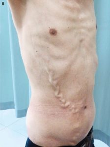
A 57-year-old man with a 5-year history of intermittent edema of the legs was admitted to the hospital because of varices in the right abdominal and chest walls (Panel A).
The varices had been gradually increasing in size and number over a period of 18 years. Abnormal results of laboratory tests included a total bilirubin level of 44.0 μmol per liter (2.6 mg per deciliter; normal range, 3.4 to 20.5 μmol per liter [0.2 to 1.2 mg per deciliter]), a serum albumin level of 28.2 g per liter (normal range, 40 to 55), and a prothrombin time of 17.1 seconds (normal range, 9.8 to 12.1). Contrast-enhanced computed tomography of the abdomen revealed a thrombus in suprahepatic and posthepatic portions of the inferior vena cava and multiple varices in the right abdominal wall. No ascites was found. Thrombosis of the inferior vena cava was further confirmed by abdominal ultrasonography and angiography of the inferior vena cava. Thus, a diagnosis of the Budd–Chiari syndrome with thrombosis of the inferior vena cava was made. Subsequent percutaneous recanalization of the inferior vena cava by means of balloon angioplasty and stent placement was successful; after the percutaneous recanalization, the patient received anticoagulation therapy for 6 months and had an excellent clinical response. At the sixth postoperative month, the abdominal-wall varices had improved markedly (Panel B).

