Face Avulsion and Degloving
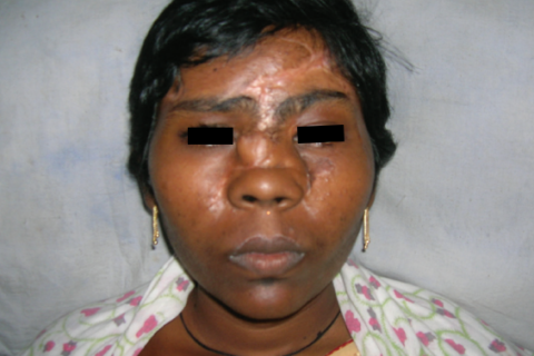
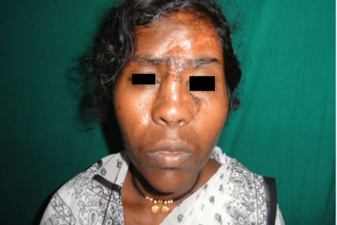
A woman presented at a hospital with a history of domestic violence involving a sickle. The attack resulted in the degloving and avulsion of the left upper hemi-face, including the nose, forehead skin, eyebrow, upper and lower eyelids, and part of the cheek. Despite the severity of the injuries, the globe of the eye and the eyesight were both intact. A thorough evaluation under anesthesia revealed a loss of the anterior cortex of the frontal sinus, and the nasal bones were also missing. The wound was contaminated with small mud particles.
The surgical team undertook a meticulous debridement of the devitalized tissue and then attempted to reposition the degloved flap and the structures to their respective positions, like a jigsaw puzzle. Meticulous suturing was done to relocate the avulsed structures to their respective positions. Medial and lateral canthopexies were done to relocate the eyelids at their respective positions under appropriate tension.
However, there was significant tissue loss in the left forehead region, over the nasal dorsum, and in cheek areas. The exposed frontal sinus was cured thoroughly to scrape all the mucosa, and the cavity was plugged with pieces of the outer cortex of the frontal sinus. A forehead flap was raised based on the right supraorbital and supratrochlear vessels and transposed medially to cover the exposed frontal sinus and exposed frontal bone. A proximally based nasolabial flap was used to cover the skin defect on the dorsum and right lateral wall of the nose. A small defect on the cheek region was split skin grafted.
The postoperative course was uneventful, and four weeks after the surgery, a costal cartilage graft was used for nasal augmentation. The cartilage was placed in an L-shaped manner for augmentation of the columella and dorsum of the nose. Thinning of the Nasolabial flap was also done in the same setting. The postoperative course was again uneventful.
A year after the trauma and two surgeries, the patient was satisfied, and there were no functional deficits. The cosmetic outcome was considered suboptimal, but the patient declined further surgery.

Fig. 2: Intra-operative picture showing canthopexy,
harvested nasolabial flap and forehead flap.
Disclaimer – Clicking on the image will open a new tab directing you to the image of the original source of this case report. Medical gore might be present.

