Giant pelvic abscess with sepsis
Case presentation
A 71 year old female presented to the emergency department complaining of right hip and flank pain that began approximately 1 month prior to arrival. Of note, she had a hysterectomy 2 years prior that was uneventful. She also stated that 15 days prior she noted redness and swelling in the same area where the pain is located. She denied fever, chills, headaches, dizziness, abdominal pain, nausea, vomiting, dysuria or any other symptoms. She also denied any trauma to the area.
Upon physical exam there was found to be a large, indurated, fluctuant mass on the right hip/flank which was spontaneously draining pus from a small opening (Fig. 1). She was diagnosed with sepsis because of her tachycardia, tachypnea, thrombocytosis, and marked leukocytosis.

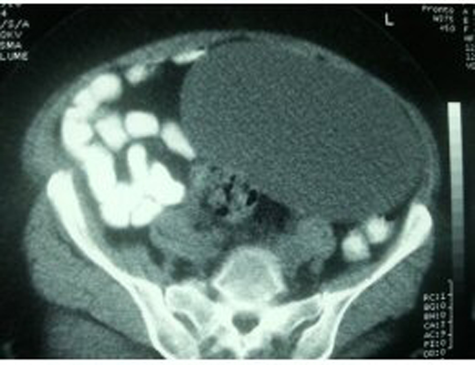
Computed tomography (CT) demonstrated a trans-fascial, multi-loculated fluid collection containing gas bubbles, extending through the right abdominal sidewall involving the retroperitoneal space, abdominal/pelvic side wall musculature, and in the subcutaneous fat compatible with abscess (Fig. 2).
A giant abscess was encountered with 3 liters of purulent fluid; intraoperative cultures were taken and sent to microbiology to identify causative organism(s). Following drainage, extensive excisional debridement was undertaken, until all necrotic tissue was removed. The wound defect was measured to be 19 cm × 20 cm × 20 cm (Fig. 3).

Vacuum assisted closure dressing (VAC) was applied to prepare the wound for a secondary examination and delayed primary closure (Fig. 4).

Post-operatively
She was taken back to the operating room 3 days later for a second look. No further wound debridement was needed. Two Jackson-Pratt drains were placed in the cavity for further drainage. According to the culture results, microbiology was not able to identify a particular predominant organism, however multiple gram-positive rods were noted and there were no anaerobes seen.
She further improved and was discharged 3 days later (hospital day 6) on oral antibiotics (as recommended by infectious disease specialist), and home health for drain management.
There were no post-operative complications noted at this time; the incision was healing well and there was no sign of infection. She was seen again 2 weeks later in clinic and no post-operative complications were noted.
Credits: Dr. Adel Elkbuli, ncbi