Prelaminated Free Flap for Auricular Reconstruction
A 42 year old woman presented with nearly complete defects of the left auricular and lateral temporal bone as a result of advanced basal-cell carcinoma. She did not want an adhesive auricular prosthesis and was not a candidate for traditional repair techniques because of trauma and irradiation damage to the postauricular skin. A prelaminated radial forearm free flap was selected for reconstruction. The term “prelamination” refers to the surgical introduction of novel tissue to an existing flap — in this case, autologous costal cartilage inserted into the patient’s forearm (Panel A).
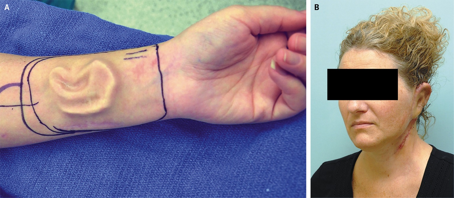
After subsequent integration and neovascularization of the added tissue, the multilayered, prelaminated flap was inset with the use of microvascular surgery to repair the defect. At follow-up 4 months after surgery, the auricular reconstruction was intact (Panel B). Potential complications of this technique include the exposure and infection of cartilage, inferior migration of the framework, poor lobule formation, distortion of the contour of the framework, and a blunted scaphoconchal angle.