Maxillofacial Injuries due to Bear Mauling
Case Report 1
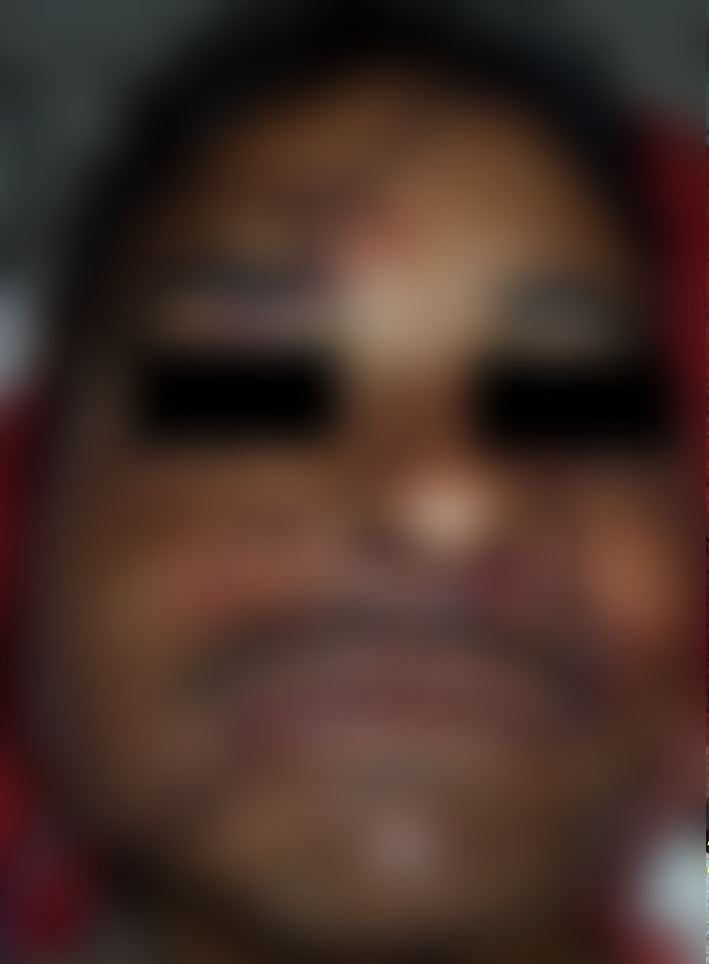
A 40-year-old male patient was presented with severe maxillofacial injuries caused by bear attack. Patient had multiple skin lacerations resulting in exposure of underlying structures (parotid gland with duct, buccinators muscle, buccal pad of fat etc.) (Fig. 1) and was bleeding profusely from nose, and oral cavity. There were degloving injuries in upper and lower vestibule, and avulsion of upper anterior teeth in addition patient had dislocated jaw, multiple fractures of maxillary bone and was delirious at presentation. CT-scan revealed that there was bilateral fracture of alveolar process of maxilla with comminution on left side, comminuted fracture of hard palate, complete fractures of all the wall of maxillary sinus bilaterally with displacement of fractured fragments into the sinuses on right side. There was bilateral fracture of nasal bone—along with fractured zygomatic arch and temporal bone on right side.
Case Report 2
A 53-year-old patient reported with soft tissue avulsion from right periorbital region, including extensive damage of nose and upper lip due to bear mauling (Fig. 1). Patient was bleeding profusely from nose and oral cavity. CT-scan revealed no bony injuries.
Management
Both the cases were treated following a basic protocol as explained below:
After maintaining the airway, breathing and circulation, bleeding was arrested by placing the anterior nasal pack with paraffin. Tetanus prophylaxis and rabies vaccine were given to the patients. After thorough surgical debridement of wound, decision was taken to close the wound primarily under local anesthesia. Intraoral layer wise closure of the wound was done with 4-0 vicryl suture. After the adequate approximation of deep structures extra oral wound closure was done using 4.0 silk sutures and a suction drain (no. 12) was placed (Figs. 2, 3). A course of broad spectrum antibiotic and anti-inflammatory was given for one week; in addition for the first case, it was decided to manage the fractured segments conservatively with intermaxillary fixation by using arch bars.


