Total Nose and Upper Lip Replantation
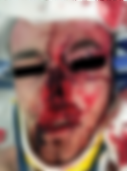
A 17 year old male presented to the emergency room 2 hours after sustaining an amputation of his nose and upper lip as he was ejected through the windshield in a motor vehicle accident. The amputated nose and upper lip had been wrapped in gauze and placed on ice at the scene of the accident. The upper lip was amputated in continuity with the nose, resulting in a 90% defect of the upper lip (Fig.1).
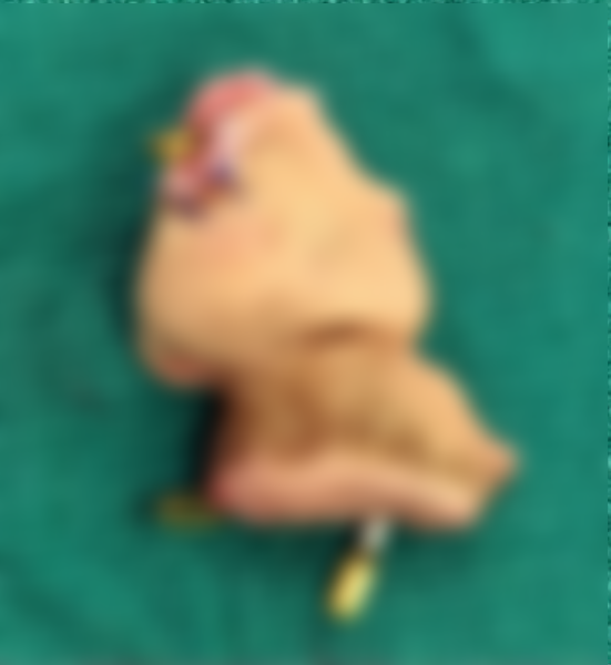
The amputated nose and upper lip segment was immediately brought to the operating room for exploration. Microsurgical clamps were used to mark the superior labial arteries (1.0 mm in diameter) bilaterally and the left dorsal nasal artery (0.5 mm) (Fig. 2).
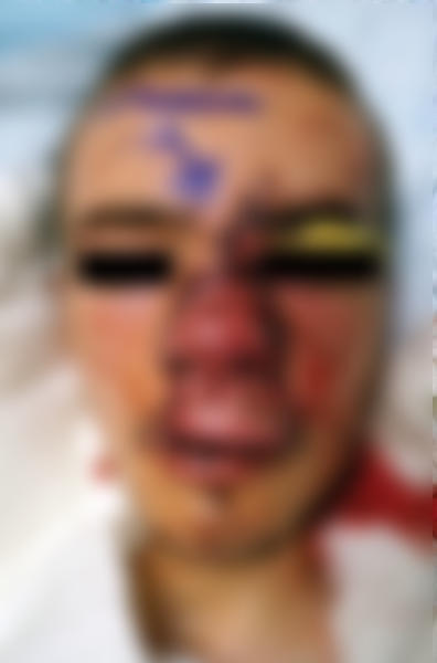
Further dissection was performed from the glabellar wound cranially to move further away from the zone of injury. A suitable 0.8 mm vein was found 3.5 cm cranially. A 4 cm interposition vein graft was harvested from the medial aspect of the right foot. As the venous anastomoses were completed, a 5,000 unit heparin bolus was given intravenously. Good venous outflow was achieved and maintained (Fig. 3)
The patient was kept on bed rest for the first 3 days with his head elevated in a warm room (26–28°C). His diet and activity level were gradually increased. He was discharged home on POD 10. He was able to quit smoking following surgery.
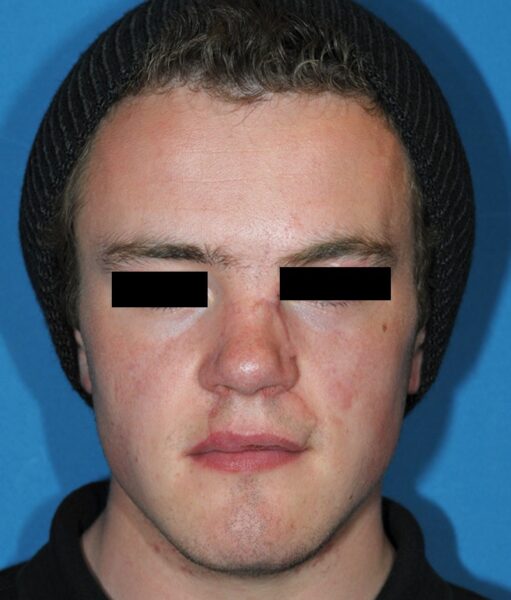
No complications occurred postoperatively. At his 18 month follow-up, the patient demonstrated excellent nasal airway patency and was very satisfied with his aesthetic result (Fig. 4)
Corresponding author. Kate Elzinga, MD, FRCSC, Abelardo Medina, MD, PhD, and Regan Guilfoyle, MD, FRCSC


