Penetrating aortic injury left untreated for 20 days: a case report
Authors: Alessia Giaquinta, Dovile Mociskyte, Giuseppe D’Arrigo, Giuseppe Barbagallo, Francesco Certo, Massimiliano Veroux & Pierfrancesco Veroux
Case presentation
A 26-year-old Libyan man was a victim of a firearm wound while he was running seeking for refuge, with a bullet penetrating his abdominal wall from the left to right side, with an entrance orifice of approximately 3 cm in diameter at the left lumbar paravertebral region. No exit wound was seen. After the assault, the victim, a clandestine refugee from Libya, spent up to 20 days crossing the Mediterranean Sea to leave his country of origin. He was finally admitted to a recovery center in Italy and then to a local hospital, in good general condition and with no signs of hemodynamic shock.
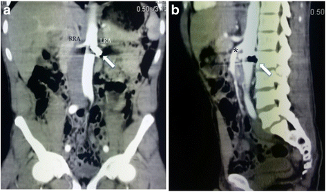
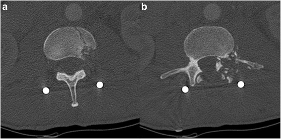
Abdominal radiography revealed the presence of a bullet located anteriorly to the second lumbar vertebra, with the tip rotated in the upright direction and fractures of the second and third lumbar vertebrae (Fig. 1). Subsequently, the patient was immediately transferred to the department of neurosurgery of our hospital. At admission, he was hemodynamically stable, with a stable hemoglobin value (10.1 g/dl), a Glasgow Coma Scale of 15, and with lumbar pain, hyposthenia, and hyperesthesia of the left lower limb. Preoperative computed tomography (CT) angiography, unexpectedly, demonstrated that the bullet penetrated partially into the aortic wall at the level of the left renal artery, without any signs of active blood loss or surrounding hematoma (Figs. 2, 3).


The patient was immediately scheduled for surgical repair of the aortic injury. The aorta was accessed retroperitoneally through a left lobotomy. The abdominal aortic segment was entirely exposed by a retrorenal access and section of the fibers of the left pillar of the diaphragm and then isolated before dealing with the bullet. At the level of the left renal artery, a bullet partially penetrating the left latero-posterior aortic wall was visualized. The bullet crossed the lumbar spine, fragmenting the second and third lumbar vertebrae, and finally penetrated the aortic wall for half of its length, creating a plug that avoided immediate life-threatening bleeding at the time of the gunshot injury.
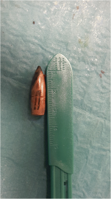
There were no signs of visceral injury or surrounding hematoma. After clamping of the aorta and the left renal artery, the bullet was clearly visible on the posterior surface of the aorta, just behind the left renal artery (Video 1). Subsequently, it was removed and the aortic lesion was repaired by a 5–0 polypropylene running suture. The extracted bullet was about 27 mm in length, with a spritzer (pointed) nose, resembling a military rifle cartridge (Fig. 4). The surgical intervention was completed by the neurosurgery team by using spinal fixation with pedicle screws at the level between the second and fourth lumbar vertebrae. The immediate postoperative course was uneventful, and computed tomography angiography performed on postoperative day 3 did not demonstrate any sign of aortic bleeding and showed good results of the spinal fixation. Consequently, the patient was discharged 6 days after the surgical procedure, without any neurological symptoms.

References
- 1.Demetriades D, Theodorou D, Murray J, Asensio JA, Cornwell EE 3rd, Velmahos G, et al. Mortality and prognostic factors in penetrating injuries of the aorta. J Trauma. 1996;40:761–3.
- 2.Trimble C. Arterial bullet embolism following thoracic gunshot wounds. Ann Surg. 1968;168:911–6.
- 3.Jaha L, Ademi B, Ismaili-Jaha V, Andreevska T. Bullet embolization to the external iliac artery after gunshot injury to the abdominal aorta: a case report. J Med Case Rep. 2011;5:354.
- 4.Carter CO, Havens JM, Robinson WP, Menard MT, Gatesa JD. Venous bullet embolism and subsequent endovascular retrieval – a case report and review of the literature. Int J Surg Case Rep. 2012;3:581–3.
- 5.Stefanopoulos PK, Hadjigeorgiou PK, Filippakis K, Gyftokostas D. Gunshot wounds: a review of ballistics related to penetrating trauma. J Acute Dis. 2014;3:178–85.
- 6.Fackler ML. Gunshot wound review. Ann Emerg Med. 1996;28:194–203.
- 7.Bellamy RF, Zajtchuk R. Conventional warfare: ballistic, blast, and burn injuries. Washington, DC: Walter Reed Army Medical Center, Office of the Surgeon General; 1991. p. 107–62.
- 8.Kneubuehl BP. Wound ballistics of bullets and fragments. In: Kneubuehl BP, Coupland RM, Rothschild MA, Thali MJ, editors. Wound ballistics: basics and applications (translation of the revised 3rd German edition). Berlin: Springer; 2011. p. 163–252.
- 9.Scott R. Pathology of injuries caused by high-velocity missiles. Clin Lab Med. 1983;3:273–94.
Original source
Copyright
Open Access This article is distributed under the terms of the Creative Commons Attribution 4.0 International License (http://creativecommons.org/licenses/by/4.0/), which permits unrestricted use, distribution, and reproduction in any medium, provided you give appropriate credit to the original author(s) and the source, provide a link to the Creative Commons license, and indicate if changes were made. The Creative Commons Public Domain Dedication waiver (http://creativecommons.org/publicdomain/zero/1.0/) applies to the data made available in this article, unless otherwise stated.