14 Kilogram Subcutaneous Lipoma
Case Report
A 54 year old man with a past medical history significant only for hypertension presented to the clinic with a large soft tissue growth on
hislower back which had been present for the past 20 years.
The patient had an unremarkable physical exam except for the large soft tissue mass over the lower back, with the maxiumum dimension measured to be 38cm (Figure 1) .

lateral view
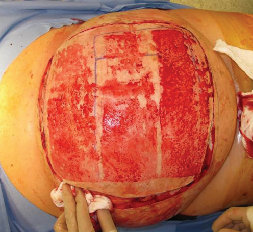
Four weeks after presentation, several surrgical teams performed a six hour operation to remove the 14 kilogram mass. After the patient was widely prepped and draped, the skin overlying the central portion of the tumor was shaved and harvested as multiple split thickess skin grafts (Figure 2).

harvest of skin grafts and initial skin- preserving
incision. The patient’s head is to the left.
Subsequently, an incision was made in the skin overlying the tumor in an
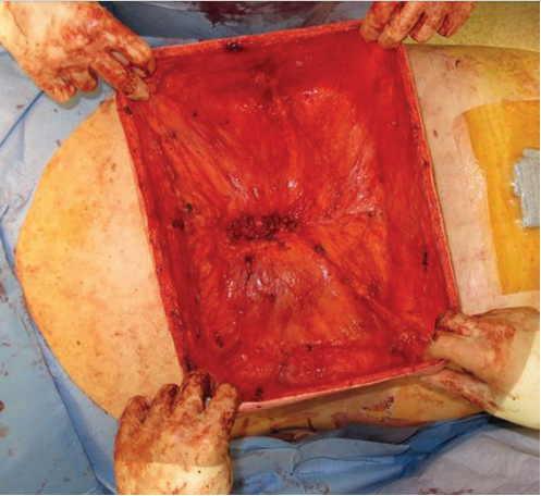
area outside the skin graft donor sites, preserving significant flaps in all dimensions to permit primary closure (Figure 3).

flaps used for reconstruction after tumor enucleation
prior to de-epithelialization and imbrication. The
patient’s head is to the left.
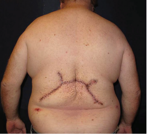
Two subcutaneous closed-suction drains were placed prior to the final closure. Postoperatively, the patient did well without complication
(Figure 4).

posterior view, demonstrating the final closure.
After a brief and uneventful hospital stay postoperatively, he was discharged home in good condition. On follow-up, his drains were
sequentially removed and the incision line has healed without problems. He has not had any evidence of recurrence or infection at six months
postoperatively.
Credit: Joseph A. Ricci, MD, The Division of Plastic Surgery,
Department of Surgery The Brigham and Women’s Hospital