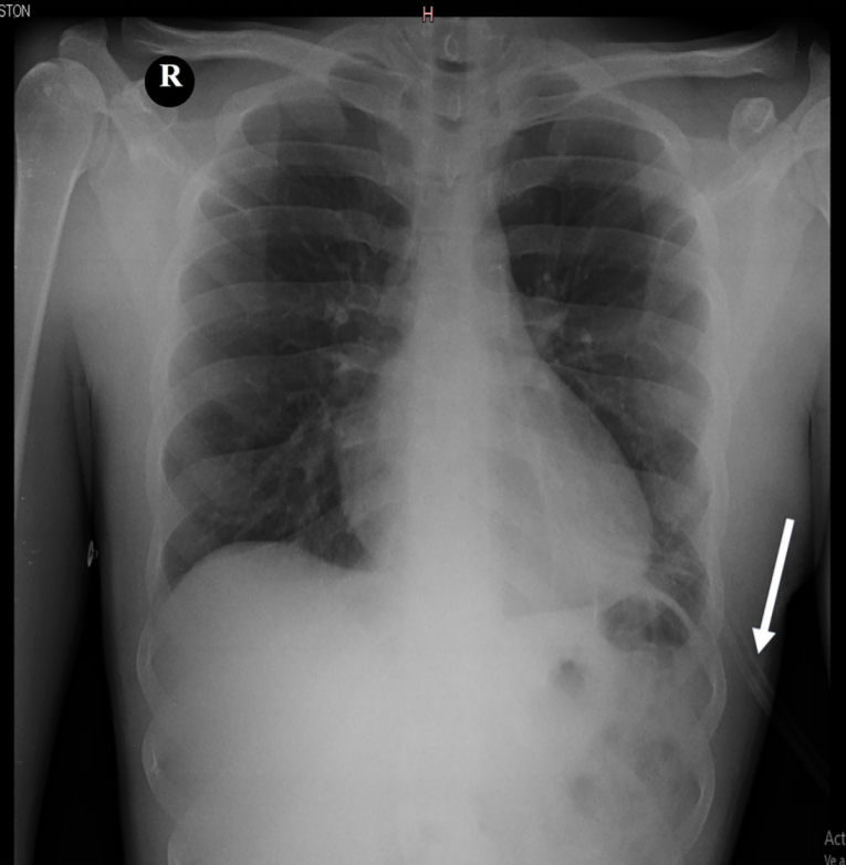
Lung Protrusion: Tragic Consequence of a Knife Assault

Lung Protrusion refers to the exposure of lung parenchyma from a defect in the chest wall. This is a case of knife assault which caused lung evisceration.
A 20-year-old presented to the Emergency Department with bilateral stab wounds in the thorax, causing lung protrusion. He also had exposed lung parenchyma on the left side. He was breathless at the time of presentation with unbearable pain on both sides of the chest wall. However, he was conscious and responding. His vitals showed low blood pressure, increased heart rate, increased respiratory rate, and normal oxygen levels. There was no other injury.
Immediate intervention
The doctors administered 1L of normal saline to the boy. Moreover, Ketoprofen and Tramadol were also given to relieve pain. To manage shortness of breath, he was provided oxygen at the rate of 6 Litres/min.
Fortunately, his blood pressure stabilized. Moreover, there was no need for blood products.
The protruded lung: Treament?
Initially, the eviscerated lung portion was covered with a sterile towel dipped in normal saline.
CT scan was ruled out. This was due to the risk of instability inside the machine. Therefore. the trauma team was called for an assessment. They evaluated the patient’s status.
Operating the wound: The final obstacle
The trauma team had advised an emergency operation. Fortunately, the eviscerated lung, the “lingula” was not necrosed. There were no other complications. Moreover, the left side showed hemothorax.
Restoration of normal anatomy
The operation was a success. Furthermore, the surgeons irrigated the thorax with normal saline. They intubated the boy after this. There was no air leakage. They suctioned the fluid out and sutured the thorax. A thoracostomy tube drained the excess secretions.

Post-operative results and follow-up
The doctors observed the boy for 12 hours. He spent this time in the general ward, not requiring any emergency intervention. After 48 hours, doctors performed a chest X-ray. It showed normal lung expansion. Furthermore, the doctors gave him painkillers and antibiotics.
The doctors followed up with the young boy. Fortunately, the wound was healing normally and he was fully functional.
Author (s)
Ferreira-Pozzi, MartínErramouspe, Pablo JoaquinFolonier, Juan CarlosPerez, Mauro PerdomoGonzález, Daniel GonzálezLaurin, Erik G.