A Rare Syndrome of Five Finger Hands and Polydactyly of the Feet: A Case Report
Ahmadreza Afshar
Case report
A 22-year old man was referred from the Welfare Organization for evaluation of his hands and feet abnormalities. The family history was negative for any congenital abnormalities. The parents were non-consanguineous. It seemed that the hands and feet deformities were symmetric. The right and the left thumbs were absent (Figs. 1 and 2). The thenar prominences were absent and the first web space was narrow on both sides. The right and left hands had five fingers. The most radial fingers were in the same plane of the other fingers and they could not be opposed to the other fingers. He had a side-to-side pinch between the most radial finger and the index finger of his hands. He was able to grasp objects with all the fingers of both hands.
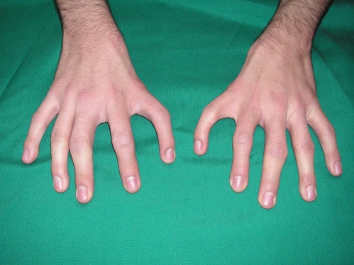
Dorsal view of the hands
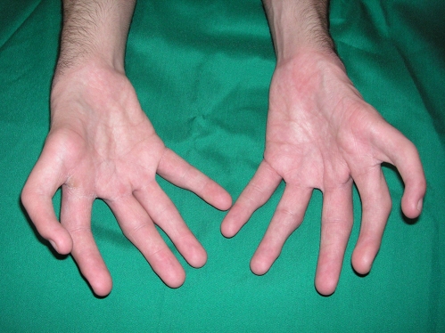
Palmar view of the hands
The arcs of wrists flexion-extension were the same on both sides and they were 70° of flexion and 45° of extension. The right elbow had a normal range of motion. The left elbow’s range of motion was 40 to 120° of flexion. The active supination-pronation arc on the right side was 100°. The active supination-pronation arc on the left side was more restricted than the right side and it was about 40°. The pronounced restricted ranges of movements on the left side might be explained by relative underdeveloped osseous structures and soft tissue abnormalities.
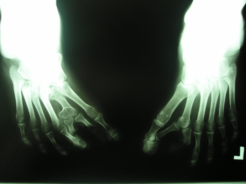
The patient had polydactyly of the feet (Fig. 3). Each foot had seven toes. Legs and the ankles appeared normal on the both sides. No chromosomal abnormality was found on karyotype.
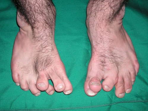
Polydactyly of the feet
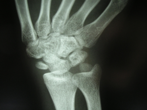
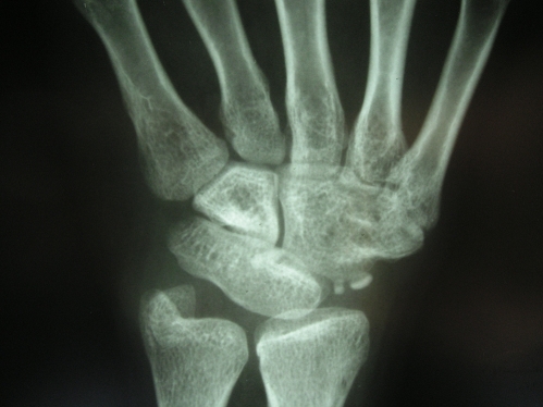
Radiographs of the hands showed that the most radial digits had a complete metacarpal and three phalanges (Fig. 4). The skeletons of the wrist joints were abnormal. Radiograph of the right wrist showed a hypoplastic distal radius and a rudimentary scaphoid. The trapezium, trapezoid and capitate bones in the distal carpal row had coalition (Fig. 5). Radiograph of the left wrist revealed a hypoplastic distal radius and a rudimentary ossicle instead of the scaphoid. The trapezium, trapezoid and capitate bones in the distal carpal row had coalition. The lunate and triquetrum were also fused (Fig. 6).
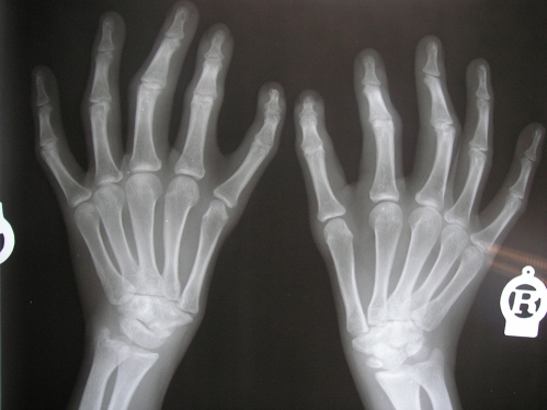
Radiography of the hands
Antro-posterior radiography of the feet
Radiography of the right wrist
Radiographs of the feet demonstrated seven digits and six metatarsals. The second and third medial digits were articulated with the second medial metatarsal at the metatarsophalangeal joints on both sides (Fig. 7).
Radiography of the left wrist
To improve the function and aesthetic of the hands’ pollicization of the most radial fingers of the hands were considered but the patient declined surgery.
Discussion
Pashayan et al reported a familial syndrome in a father and his daughter that the daughter had bilateral five finger hands, absent thumbs, bilateral tibial aplasia, and preaxial polydactyly of the feet. Her father hands were identical to his affected daughter with five finger hands and no thumbs [3]. Borg et al have reported a case of tetramelic mirror-image polydactyly with unilateral tibial hypoplasia [4]. Lamb et al reported a kindred of 15 individuals in five generation with absent thumbs, five finger hands, partial or complete tibial absence and preaxial polydactyly [5]. The symmetric abnormalities in the hands and feet of the above reports suggest an autosomal dominant gene cause to the development of the four limbs. However, the authors of the above reports did not point to the exact genetic abnormality [3–5]. Kondoh et al and Kim et al have suggested a novel gene disrupted at a 14q13 breakpoint of t(2;14) which produces mirror-image polydactyly of hands and feet [6, 7]. However, the genetic abnormality might have variable expressivity to produce absent thumb, preaxial polydactyly of the hands and feet, and with or without varying degree of tibia aplsia
The embryonic limb buds development through proximodistal, radioulnar and dorsoventral axes. Proximodistal development depends on the apical ectodermal ridge (AER) signals. Radioulnar development depends on the morphogenic signals from the zone of polarizing activity (ZPA) located at the posterior border of the limb. AER and ZPA signals constitute a reciprocal feedback loop [8].
One explanation for the abnormalities of the limbs in this case might be that the genetic abnormality altered the AER and ZPA the activities [8]. Failure of the signals to reach the preaxial side from the AER leads to abort the development on the preaxial side of the feet and produce absence of the thumbs in the hands [8]. Ectopic ZPA morphogenic signals on the preaxial side of the limbs lead to produce five-fingered hands and mirror feet anomalies [8]. In hand ectopic ZPA signals on the radial side of the index finger produce finger duplication [9].
Al-Qattan et al have indicated that the phenotype of the mirror hand deformity is variable and patients may have absent thumbs, preaxial polydactyly and a hypoplastic radius. The presence of hypoplastic radius in the current case supports that the case may well be a variant of mirror hand [1].
In this case, the anomalies might be a rare syndrome of atypical mirror hands and mirror feet. This case is presented because of its rare occurrence and each reported case will hopefully bring more insight to the congenital anomalies.
References
References
- Al-Qattan MM, Al-Thunayan A, Cordier M, Nandagopal N, Pikaanen J. Classification of the mirror hand—Multiple hand spectrum. J Hand Surg. 1998;23B:534–536. [PubMed] [Google Scholar]
- Verghese R, Shah H, Rebello G, Joseph B. Pre-axial mirror polydactyly associated with tibial deficiency: a study of the patterns of skeletal anomalies of the foot and leg. J Child Orthop. 2007;1:49–54. doi: 10.1007/s11832-006-0001-5. [PMC free article] [PubMed] [CrossRef] [Google Scholar]
- Pashayan H, Fraser FC, McIntyre JM, Dunbar JS. Bilateral aplasia of the tibia, polydactyl and absent thumb in father and daughter. J Bone Joint Surg. 1971;53B:495–499. [PubMed] [Google Scholar]
- Borg DH, Roermund PM, Kon M. A sporadic case of tetramelic mirror-image polydactyly and unilateral tibial hypoplasia without associated anomalies. J Hand Surg. 1999;24B:482–485. [PubMed] [Google Scholar]
- Lamb DW, Wynne-Davis R, Whitmore JM. Five-fingered hand associated with partial or complete tibial absence and pre-axial polydactyly. J Bone Joint Surg. 1983;65B:60–64. [PubMed] [Google Scholar]
- Kondoh S, Sugawara H, Harada N, et al. A novel gene is disrupted at a 14q13 breakpoint of t(2;14) in a patient with mirror-image polydactyly of hands and feet. J Hum Genet. 2002;47:137–139. doi: 10.1007/s100380200015. [PubMed] [CrossRef] [Google Scholar]
- Kim KC, Wakui K, Yamagishi A, et al. Tetramelic mirror-image polydactyly and a de novo balanced translocation between 2p23.3 and 14q13. Am J Med Genet. 1997;68:70–73. doi: 10.1002/(SICI)1096-8628(19970110)68:1<70::AID-AJMG13>3.0.CO;2-L. [PubMed] [CrossRef] [Google Scholar]
- Oberg KC, Feenstra JM, Manske PR, Tonkin MA. Developmental biology and classification of congenital anomalies of the hand and upper extremity. J Hand Surg. 2010;35A:2066–2076. [PubMed] [Google Scholar]
- Al-Qattan MM, Yang Y, Kozin SH. Embryology of the upper limb. J Hand Surg. 2009;34A:1340–1350. [PubMed] [Google Scholar]
Articles from Journal of Hand and Microsurgery are provided here courtesy of Thieme Medical Publishers
Author information
Department of Orthopaedics, Imam Khomeini Hospital, Urmia University of Medical Sciences, Modaress Street, Ershad Boulevard, Urmia, Iran 57157-81351
Ahmadreza Afshar, Phone: +98-912-3131556, Fax: +98-441-3469939
Copyright and License
Copyright © Society of the Hand & Microsurgeons of India 2011