Avulsed tissue after dog bite
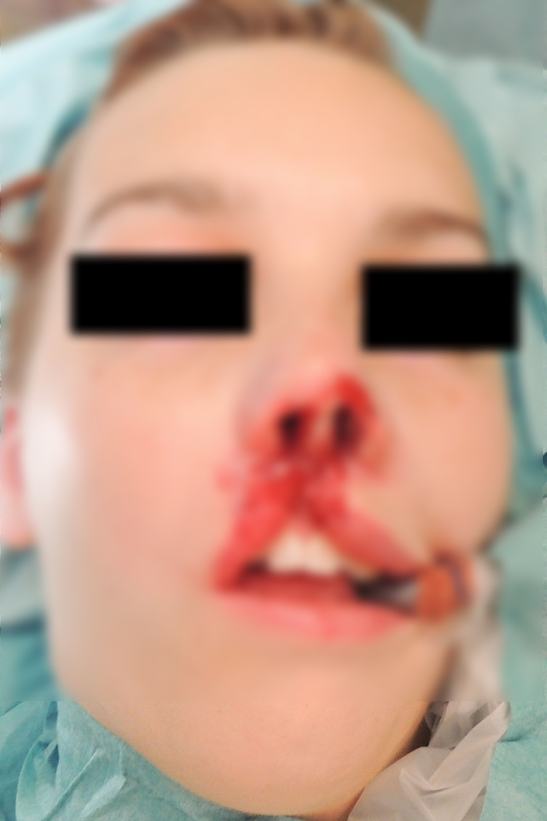
An 18-year-old otherwise healthy woman was attacked by a family dog and suffered a bite to the central part of the face (Fig 1).

The injury on arrival at the trauma centre
The avulsed tissue was conserved in a plastic bag within another plastic bag with iced water. It consisted of the nose tip, the entire left ala, half of the right ala and approximately 3cm of the upper lip including the philtrum (Fig 2).

Avulsed tissue
However, three initial attempts resulted in no blood flow. Only after a wrist vein was used for a vein graft was a satisfactory (albeit not optimal) blood supply achieved (Fig 3). The patient was admitted to the intensive care unit, and remained intubated and sedated for just over 24 hours.

After replantation
On day 4 after the trauma, the flap grew increasingly darker. Nevertheless, a conservative approach was taken and after ten days, the patient was discharged while awaiting demarcation of the flap (Fig 4).

Early demarcation
One month later, the flap was deemed to be non-vital, with dry necrosis of the skin (Fig 5). Expecting a large defect and reconstruction with both an Abbe flap and forehead flap, the patient underwent surgical debridement and although the skin was clearly not viable, healthy tissue was surprisingly found immediately underneath (Fig 6).

Skin necrosis
After removal of superficial skin necrosis
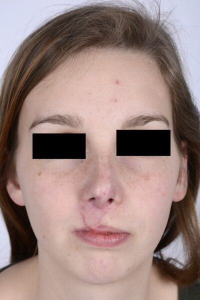
After transposing the orbicularis muscle to give the lip some central ‘fullness’, the defect was covered with a full-thickness postauricular skin graft (Fig 7). The skin graft took 100% and follow-up review has shown an acceptable cosmetic result (Fig 8) but further surgery might be indicated to optimise the result.

Full-thickness skin graft
Credit: CORRESPONDENCE To Dr. Jògvan øregaard