Carotid–Cavernous Sinus Fistula
A 55 year old woman with a long history of hypertension presented to the hospital with a 1-day history of periorbital discomfort, inferior chemosis, and conjunctival injection of the left eye (Panel A).

She reported having a 2-year history of episodic headache and pulsatile tinnitus in the left ear. Examination of the left eye revealed a visual acuity of 20/60, a relative afferent pupillary defect, exophthalmos (with 6 mm of protrusion), and an elevated intraocular pressure (48 mm Hg). Examination of the right eye was unremarkable. Contrast-enhanced computed tomography of the orbit showed a dilated left superior ophthalmic vein (Panel B, arrow), suggesting the presence of a carotid–cavernous sinus fistula.
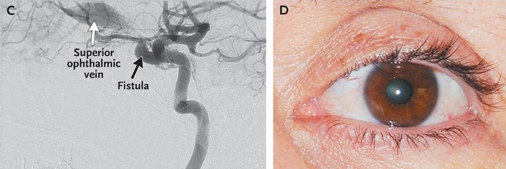
Angiography confirmed that the internal carotid artery was connected to the cavernous sinus by a large fistula (Panel C, black arrow). Dilatation of the superior ophthalmic vein (Panel B, arrow; Panel C, white arrow) is a key feature of this condition and results from the transmission of arterial pressures to the venous system. After embolization of the fistula with the use of seven platinum coils, the patient had complete resolution of the relative afferent pupillary defect, elevated intraocular pressure, and exophthalmos, and her visual acuity returned to normal (Panel D).