Dilated cardiomyopathy

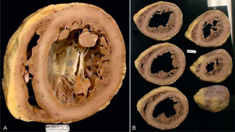
Idiopathic dilated cardiomyopathy. Views of the heart in a 61-year-old woman who had had evidence of chronic heart failure since age 51 years. She did reasonably well on medical therapy until age 59 years, when the heart failure worsened considerably and an implantable cardiac defibrillator was inserted. During the 2 years before transplant, the heart failure progressively worsened, and the left ventricular ejection fraction fell to a low of approximately 10%. She never had chest pain. Earlier in life she had had several children. (a) Basal portion of heart exposing well the mitral valve. The left ventricle is greatly enlarged measuring up to 7.5 cm. No foci of fibrosis or necrosis are seen in the ventricular walls. (b) Views of the ventricles caudal to the mitral valve, again showing considerable left ventricular cavity dilatation.

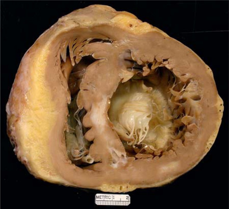
Hypertrophic cardiomyopathy. Heart in a 41-year-old man in whom hypertrophic cardiomyopathy was diagnosed when he was 6 years of age. At age 26 years, atrial fibrillation appeared and during the next 15 years he was cardioverted 98 times. He eventually developed complete heart block, and a pacemaker was inserted. At age 30 years, an intracardiac defibrillator was implanted, and at the same time an alcohol septal ablation procedure was performed. Nevertheless, evidence of heart failure persisted and a heart transplant was performed. View of the basal portion exposing both tricuspid and mitral valves. The left ventricular cavity is quite dilated. The focal scar in the ventricular septum may be the result of the alcohol septal ablation. Another small scar is present in the posterior wall of left ventricle. The thickness of the ventricular septum is greater than that of the left ventricular free wall.