Giant Lipoma of Posterior Neck with Bleeding Decubitus Ulcer: A Rare Entity
Satyajeet Verma, Manish Varma, Sanjay Kala, and RK Singh
CASE REPORT
A 68-year-old male presented to our surgical outpatient department with a huge lump at the back of his neck for the last 11 years. There was an ulcer at the top of the lump for the last 9 months. Episodic bleeding was also reported from the ulcer for the last 6 months.
On local examination of the neck, there was a 22 cm × 12 cm-sized swelling at the posterior side of the neck [Figure 1]. It was nontender and soft to firm in consistency. There was a 6 cm × 5.5 cm decubitus ulcer at the top of the lump. Dilated veins were present in the skin around the decubitus ulcer. There was no regional lymphadenopathy.

Figure 1
Photograph of the patient showing giant lipoma at the posterior triangle
Fine needle aspiration revealed mature lipocytes indicative of lipomatous lesion. Contrast Enhanced Computed Tomography Scan of the neck revealed a giant subcutaneous lipoma at the posterior triangle without septations. There was no communication with the spinal cord.

Figure 2
Operative photograph showing lump excision with raised upper and lower skin flaps
After intubation with general anaesthesia, the patient was positioned in a prone position. An elliptical transverse incision of 12 cm was given at around the base of the lump. The superior and inferior skin flaps were raised [Figure 2]. Separation of lipoma from the surrounding tissues was easy and was performed with sharp and blunt dissections. Five to six large feeding vessels were also ligated to isolate the lipoma. The redundant skin was removed and the upper and lower skin flaps were stitched together with Silk-3-0 after securing haemostasis and placing a suction drain.
The resected mass was 2.2 kg in weight and 22 cm × 12 cm in diameter [Figure 3]. The postoperative period was uneventful. The drain was removed after 3 days and the patient was discharged on the 9th postoperative day.
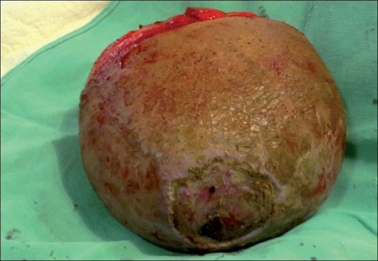
Excised lipoma with decubitus ulcer
Histopathological analysis of the resected mass revealed mature, proliferative lipocytes with no cellular atypia, and it was diagnosed as benign giant lipoma.