Giant right coronary artery aneurysm- case report and literature review
Neerod K Jha,1Husam Z Ouda,2Javed A Khan,1Gregory P Eising,1 and Norbert Augustin1
Case presentation
A 54 years-old-hypertensive male patient was presented to us with history of recent inferior wall myocardial infarction (MI) which was managed with medical treatment in the referring hospital. On clinical examination, there was a 3/6 ejection systolic murmur along the left lower sternal border. Electrocardiogram was consistent with inferior MI. The 2-D echocardiogram revealed a large cystic mass adjacent to the right atrium. Coronary angiography revealed significant coronary artery disease in the proximal left anterior descending artery (LAD) and a giant aneurysm of middle segment of right coronary artery (RCA). There was a mild ectatic segment in the proximal circumflex coronary artery, as well.
Patient underwent successful resection of giant aneurysm of RCA under cardiopulmonary bypass (CPB) via median sternotomy. Proximal and distal communications of RCA were ligated from within the aneurysmal sac and then coronary artery bypass graft surgery (CABG) was performed using right internal mammary artery to the distal RCA and left internal mammary artery to the LAD.
The aneurysmal sac was found to be 12 × 9 × 1 cms in dimension, occupying the entire right atrioventricular groove and displacing the right atrium (Figure (Figure11 &2). There was no luminal thrombus or calcification. Histopathology of excised aneurysm had shown widespread myxoid degeneration in the media, focal necrosis, atherosclerosis and fibrosis of the medial muscles.
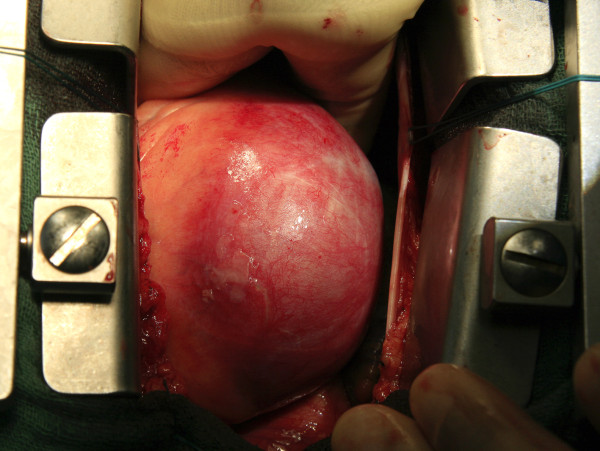
Operative photograph showing a giant aneurysm of the right coronary artery.
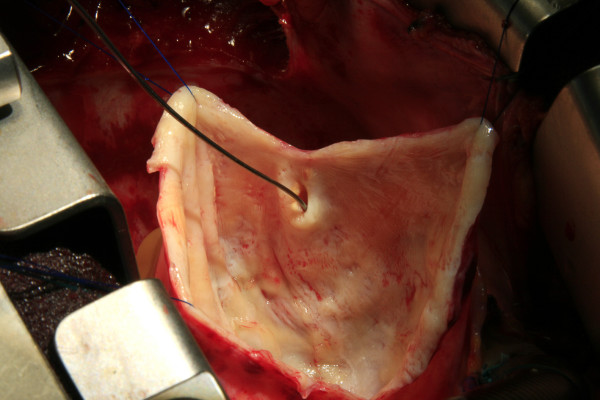
Operative photograph showing inside view of the aneurysmal sac. The tip of probe is within the proximal communication of right coronary artery.
References
- Nichols L, Lagana S, Parwani A. Coronary artery aneurysm: a review and hypothesis regarding etiology. Arch Pathol Lab Med. 2008;132:823–28. [PubMed] [Google Scholar]
- Nobrega TP, Klodas E, Breen JF, Liggett SP, Higano ST, Reeder GS. Giant coronary artery aneurysms and myocardial infarction in a patient with systemic lupus erythematosus. Catheterization and Cardiovascular Diagnosis. 1998;39:75–79. doi: 10.1002/(SICI)1097-0304(199609)39:1<75::AID-CCD16>3.0.CO;2-N. [PubMed] [CrossRef] [Google Scholar]
- Channon KM, Wadsworth S, Bashir Y. Giant coronary aneurysm presenting as a mediatinal mass. Am J Cardiol. 1998;82:1307–8. doi: 10.1016/S0002-9149(98)00626-2. [PubMed] [CrossRef] [Google Scholar]
- Yu WQ, Shou HS, Tao QS. Surgical treatment of giant coronary artery aneurysm. Asian Cardiovasc Thorac Ann. 2001;9:215–7. [Google Scholar]
- Konen E, Feinberg MS, Morag B, Guetta V, Shinfeld A, Smolinsky A, Rozenman J. Giant right coronary aneurysm-CT angiographic and echocardiographic findings. AJR. 2001;177:689–91. [PubMed] [Google Scholar]
- Hao WR, Chen FC, Kao PF, Hsieh MH, Chen YJ, Chan P. Adult giant coronary artery aneurysm-a case report and literature review. Acta Cardiol Sin. 2004;20:187–90. [Google Scholar]
- Banerjee P, Houghton T, Walters M, Kaye GC. Giant right coronary artery aneurysm presenting as a mediastinal mass. Heart. 2004;90:e50. doi: 10.1136/hrt.2002.002519. [PMC free article] [PubMed] [CrossRef] [Google Scholar]
- Grandmougin D, Croisille P, Robin C, Pacoch M, Barral X. Giant coronary aneurysm mimicking a compressive cardiac tumor: imaging features and operative strategy. Cardiovasc Pathol. 2005;14:272–5. doi: 10.1016/j.carpath.2005.04.001. [PubMed] [CrossRef] [Google Scholar]
- McGlinchey PG, Maynard SJ, Graham AN, Roberts MJD, Khan MM. Giant aneurysm of the right coronary artery compressing the right heart. Circulation. 2005;112:e66–e67. doi: 10.1161/CIRCULATIONAHA.104.496224. [PubMed] [CrossRef] [Google Scholar]
- Dianyuan L, Wu Q, Sun L, Song Y, Wang W, Pan S, Luo G, Liu Y, Qi Z, Tao T, Sun JZ, Hu S. Surgical treatment of giant coronary artery aneurysm. J Thorac Cardiovasc Surg. 2005;130:817–21. doi: 10.1016/j.jtcvs.2005.04.004. [PubMed] [CrossRef] [Google Scholar]
- Shakir D, Carr C, Suwaidi J. Giant coronary artery aneurysm and severe mitral regurgitation in patients with familial hypercholesterolemia-case report and review of the literature. Int J Angiology. 2005;14:249–52. doi: 10.1007/s00547-005-2000-7. [CrossRef] [Google Scholar]
- Augustin N, Wessely R, Porner M, Schomig A, Lange R. Giant coronary aneurysm obstructing the right heart. Lancet. 2006;368:386. doi: 10.1016/S0140-6736(06)69112-5. [PubMed] [CrossRef] [Google Scholar]
- Kumar G, Karon BL, Edwards WD, Puga FJ, Klarich KW. Giant coronary artery aneurysm causing superior vena cava syndrome and congestive heart failure. Am J Cardiol. 2006;98:986–88. doi: 10.1016/j.amjcard.2006.04.047. [PubMed] [CrossRef] [Google Scholar]
- Manghat NE, Hughes GJM, Cox ID, Roobottom CA. Giant coronary artery aneurysm secondary to Kawasaki disease: diagnosis in an adult by multi-detector row CT coronary angiography. BJR. 2006;79:e133–136. doi: 10.1259/bjr/16077689. [PubMed] [CrossRef] [Google Scholar]
- Jindal RK, George R, Singh B. Giant coronary aneurysm following drug-eluting stent implantation presenting as fever of unknown origin. J Invasive Cardiol . 2007;19:313–314. [PubMed] [Google Scholar]
- Takano MI, Oikawa M, Yamaki T, Yamaguchi O, Nakazato K, Ohsugi T, Kobayashi A, Watanabe M, Yaoita H, Yukio M. A case of recurrent myocardial infarction caused by a giant right coronary artery aneurysm. J Am Soc Echocardiogr. 2007;20:1318.e5–1318.e8. doi: 10.1016/j.echo.2007.04.008. [PubMed] [CrossRef] [Google Scholar]
- Blank R, Haager PK, Maeder M, Genoni M, Rickli H. Giant right coronary artery aneurysm. Ann Thorac Surg. 2007;84:1740–42. doi: 10.1016/j.athoracsur.2007.05.006. [PubMed] [CrossRef] [Google Scholar]
- Malero EA, Martin YD, Janelle GM, Peng YG. An unusual giant right coronary artery aneurysm resembles an intracardiac mass. Anesth Analg. 2008;107:1161–62. doi: 10.1213/ane.0b013e318181f74f. [PubMed] [CrossRef] [Google Scholar]
- Vlachou PA, Mulcahy K, Adair W. Giant coronary artery aneurysm: an unusual cause of a mediastinal mass. Eur Radiol . 2008;18:3007–3009. doi: 10.1007/s00330-008-1010-1. [PubMed] [CrossRef] [Google Scholar]
- Eshtehardi P, Cook S, Moarof I, Triller HJ, Windecker S. Giant coronary artery aneurysm. Circulation: Cardiovascular Interventions. 2008;1:85–86. doi: 10.1161/CIRCINTERVENTIONS.107.763656. [PubMed] [CrossRef] [Google Scholar]
- Matsubayashi K, Asai T, Nishimura O, Kinoshita T, Ikegami H, Kambara A, Suzuki T. Giant coronary artery aneurysm in the left main coronary artery: a novel surgical procedure. Ann Thorac Surg. 2008;85:2130–32. doi: 10.1016/j.athoracsur.2007.12.052. [PubMed] [CrossRef] [Google Scholar]
- Kanaan S, Baker C, Starnes V. resection of giant coronary artery aneurysm in a Takayasu’s arteritis patient. Ann Thorac Surg. 2008;85:1795–96. doi: 10.1016/j.athoracsur.2007.10.053. [PubMed] [CrossRef] [Google Scholar]
- Sharma J, Kanei Y, Kwan TW. A case of giant coronary artery aneurysm after placement of a heparin-coated stent. J Invasive Cardiol. 2009;21:e22–23. [PubMed] [Google Scholar]
- Pahlavan PS, Niroomand F. Coronary artery aneurysm: a review. Clin Cardiol. 2006;29:439–43. doi: 10.1002/clc.4960291005. [PMC free article] [PubMed] [CrossRef] [Google Scholar]
Articles from Journal of Cardiothoracic Surgery are provided here courtesy of BioMed Central