Maxillofacial Injuries after a Bear Attack
Case report
A 55 year old male farmer from Daporijo, Arunachal Pradesh, was referred to our hospital in September 28, 2015, with history of bear mauling. He went to the forest to hunt and tame wild bovines (Bos frontalis). He unexpectedly came across a bear (Ursus thibetanus). The bear attacked his upper body as he tried to defend himself, as a result of which, he sustained multiple soft-tissue injuries on his arms, scalp, and face.
He had a scalp wound in the occipital region of 10 cm × 12 cm size [Figure 1]. Facial wound approximately 15 cm long extends from the right lateral forehead to 3 cm superior from the right mandibular angle and medially across the upper and lower eyelid till the bridge of the nose [Figure 2]. His right orbital cavity was also involved in the injury, and the right orbit appeared to be avulsed.

He sustained right-side partial facial nerve and maxillary nerve injury along with loss of vision. He also sustained multiple fractures of the facial bones.

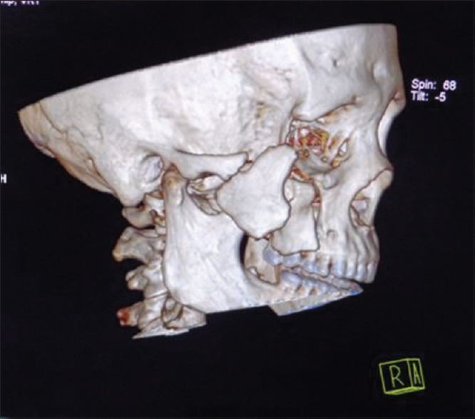
Four days after the incident, the patient was operated. The fracture was treated with open reduction and internal fixation with 1.5-mm titanium plates and screws. The zygoma was fractured from maxilla, temporal, and frontal bones into two large fragments. To align the fragments into position, two titanium screws were drilled into the fragments such that their placement does not hamper plate fixation [Figure 4].

Ophthalmic surgeon performed soft-tissue reconstruction of the upper and lower eyelids. Traction sutures were placed from the upper eyelid downward to improve eyelid closure postoperatively. The wound was approximated in layers with resorbable sutures [Figure 5].

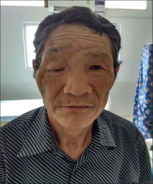
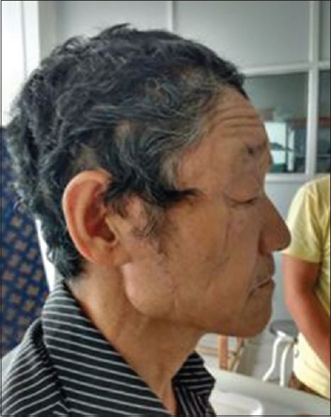
After 2 weeks, postsurgical complication of salivary fistula was noted in the parotid region. Antisialagogues were administered, and the patient was closely observed for 2 weeks. As the result was not satisfactory, the patient had to undergo superficial parotidectomy under general anesthesia. Follow-up was done for 1 year. Facial nerve injury was noticeable in the upper eyelid region and during smiling. After 8 months, the patient showed fair amount of improvement from facial nerve paralysis [Figures 6-7 ].
Credit: Dr. Mohammed Imran, ncbi