Midcarpal amputation of the hand
Case Report
A 30-year-old cheesemaker presented in their emergency room with a severe mutilating avulsion injury of the hand and forearm. His right adominant hand was pulled in a cheese cutting machine with six circular saw blades working in opposition to each other. He sustained a midcarpal amputation of the hand, a complete avulsion of the long fingers, a splitting of the forearm with extended palmar soft tissue flap, dorsal extensive soft tissue defect with laceration of the extensor muscles and an open ulnar shaft fracture, defined as third grade (IIIB) according to the classification of Gustilo and Anderson (Figure 1).

Midcarpal amputation of the hand, complete avulsion of the long fingers till midcarpal with extensive soft tissue defect.
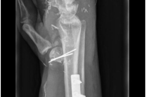
The completely amputated thumb, separated at the level of the metacarpal head, was the only finger with enough good soft tissue and bone remaining to use for reconstruction as a functional finger. In the first operation after debridement and osteosynthesis of the ulna, the thumb was pinned to a small decorticated area five centimetres (cm) proximal to the radial styloid in sense of a Vilkki-Procedure (Figure 2). In order to cover the large soft tissue defect, while in addition simultaneously reconnecting a palmar artery of the thumb to the radial artery and a dorsal vein to one in the forearm, an anterolateral thigh (ALT) flap was used as an emergency free flow-through flap (Figure 3).

The thumb was fixed to the radius in sense of a Vilkki-Procedure.

The anterolateral thigh flap.
In the second step four months later, after good healing of soft tissue had occurred, a tendon and nerve reconstruction for the thumb was performed. Initially, a neuroma at the end of the median nerve was resected. The gap between the distal end of the median nerve and the proximal end of the palmar nerve of the thumb was 8 cm. Therefore, a sural nerve graft was used to restore sensitivity (Figure 4).

The resensibilisation with a sural nerve graft.
For motorisation of the thumb, a direct suture of the flexor pollicis longus to restore flexion function was performed. Simultaneously, to accomplish the extensor function, the extensor carpi radialis longus was connected with extensor pollicis longus by a plantaris longus tendon graft (Figure 5). Subsequent to the operation, an intensive occupational therapy for mobilisation and resensibilisation of the thumb was carried out for several weeks.

The motorisation of extensor function with a plantaris longus tendon graft.
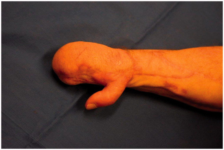
The thumb had an astonishing good function after the therapy. We measured a pinch grip of five pounds and a two-point discrimination of 10 mm at the pulp of the thumb, while there is a small area, where the sensation is felt on the amputated index finger. It is supposed as a miscellaneous sensation of median nerve. With the gripping ability of the thumb the hand can be utilised for many functions required for daily life. One year after the operation, he was able to return to work in a bigger factory and a more modern workplace with highly automated machinery (Figure 6).

Result 1 year after surgery good soft tissue conditions.
Author information
aDepartment of Hand and Plastic Surgery, Luzerner Kantonsspital, Lucerne, Switzerland,
CONTACT Dominique Merky, moc.liamg@ykremod, Department of Hand and Plastic Surgery, Luzerner Kantonsspital, LucerneSwitzerland
Copyright
Copyright © 2017 The Author(s). Published by Informa UK Limited, trading as Taylor & Francis Group.