Necrotizing fasciitis and meningitis due to Streptococcus pneumoniae
A 68-year-old Caucasian female was admitted to our clinic, with suspected deep venous thrombosis, presenting with pain in her left calf that had lasted for 3 weeks starting after a very painful nocturnal leg cramp, and worsened over the last few days. At the time of admission, she had also experienced 5 days of malaise and loss of appetite and in addition 1 day of headache and lower back pain. The patient had been coughing a lot ever since recovering from a confirmed Influenza B upper airway infection 3 months earlier. The patient received antihypertensive medication but had no other known chronical medical conditions.
At the time of admission, she was fully awake with a Glasgow Coma Scale (GCS) of 15. She was afebrile with a temperature of 36.3 °C (97.3 F) and had no neck stiffness. She presented with a well-defined erythema on her left calf (Fig. 1) and any touch or movement of her left knee, ankle or calf was very painful. There were no clinical signs of intra-articular fluid in the knee.
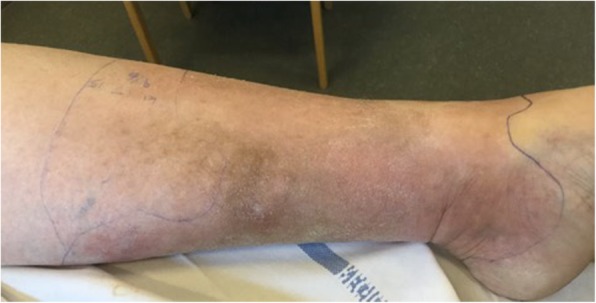
The patient’s left leg before the operation
Her blood tests showed a very high level of C-reactive protein (CRP) of 450 mg/L (normal range below 10 mg/L) and D-dimer of 18 FEU/L (normal range below 0.5 FEU/L). Her LRINEC score was 5 points (4 points from CRP 450 mg/L and 1 point from hemoglobin 12.9 g/dL (normal range between 12.1 g/dL – 15.1 g/dL)). Treatment with cefuroxime was initiated due to suspected infection of unknown origin. A CT scan of the abdomen and thorax performed due to suspected lung embolism showed discrete bilateral atelectatic areas of the lungs, but otherwise normal findings.
Four hours after admission the patient had developed an altered mental state with GCS of 9 (Eyes 2, Verbal 2 and Motor 5), neck stiffness and a fever with a temperature of 38.8 °C (102 F). Due to suspected meningitis a spinal tap was performed and empiric treatment against meningitis was initiated with Dexamethasone, Ceftriaxone and Ampicillin. The tests of the cerebrospinal fluid (CSF) showed signs of meningitis with a white blood cell count (WBC) of 50 cells/μL that were 81% granulocytes, very high lactate of 17 mmol/L, very low glucose of 0.3 mmol/L and very high protein level above 6 g/L. Initial microscopy showed Gram-positive diplococci. A CT-scan of the patient’s brain was performed and showed no abnormalities.
Eight hours after admission the patient showed a slight decline in GCS of 8 (Eyes 2, Verbal 2 and Motor 4). However, she still showed a disproportionally strong pain reaction to any touch of the left calf. The erythema had not spread since admission to the hospital, but over the next 2 h a spread of about 5 cm proximally was observed. Due to the progression of the erythema together with the strong pain reaction despite a decline in mental state, a diagnostic incision of the tissue was performed in local analgesia. The subcutis was found to be loose from the fascia and the tissues looked nonvital. Antibiotic treatment was changed to Meropenem, Clindamycin and Ciprofloxacin against NF due to national guidelines. An acute surgical débridement was performed (Fig. 2). The patient was transferred to a hospital specialized in the treatment of patients with NF, including specialized surgical care, intensive care, and hyperbaric oxygen treatment. Reinspection of the tissue in the left leg was performed with no findings of necrotic tissue. Inspection of the leg was afterwards performed routinely once daily with normal findings.

The patient’s left leg after the operation
Over the next days, S. pneumoniae was cultured both in the CSF and in 4 out of 4 blood cultures from the patient. The serotype of the bacteria was determined as 9 N. No bacteria were found by culturing the tissue from the leg, but 16S-PCR of the tissue was performed, which also showed S. pneumoniae. The 16S-PCR was performed at a department of clinical microbiology at a big university hospital. It was performed from a commercially available MicroSEQ kit with a fragment size of 500 base pairs. The cultured S. pneumoniae were susceptible to all antibiotics that were tested, including benzylpenicillin. No other bacteria were found. Therefore, antibiotic treatment was switched to benzylpenicillin on day 3 after admission.
The patient subsequently underwent reconstructive surgery with a split skin graft (Fig. 3) and was discharged after 51 days of hospitalization, including in departments of intensive care, infectious diseases and plastic surgery.

The patient’s left leg after reconstructive surgery
The patient later underwent different tests for immunodeficiency. Immunoglobulins, HIV-test and differential WBC count were all normal. The patient was not vaccinated against S. pneumoniae prior to her admission. However, she has been afterwards and serotype 9 N is included in the vaccine.
Author
1Department of Infectious Diseases, Copenhagen University Hospital, Hvidovre, Denmark
2Department of Endocrinology, Copenhagen University Hospital, Hvidovre, Denmark
3Department of Orthopedics, Copenhagen University Hospital, Hvidovre, Denmark
4Department of Infectious Diseases, Copenhagen University Hospital, Rigshospitalet, Denmark
5Department of Intensive Care, Copenhagen University Hospital, Rigshospitalet, DenmarkNichlas Hovmand