Neutrophilic Dermatosis
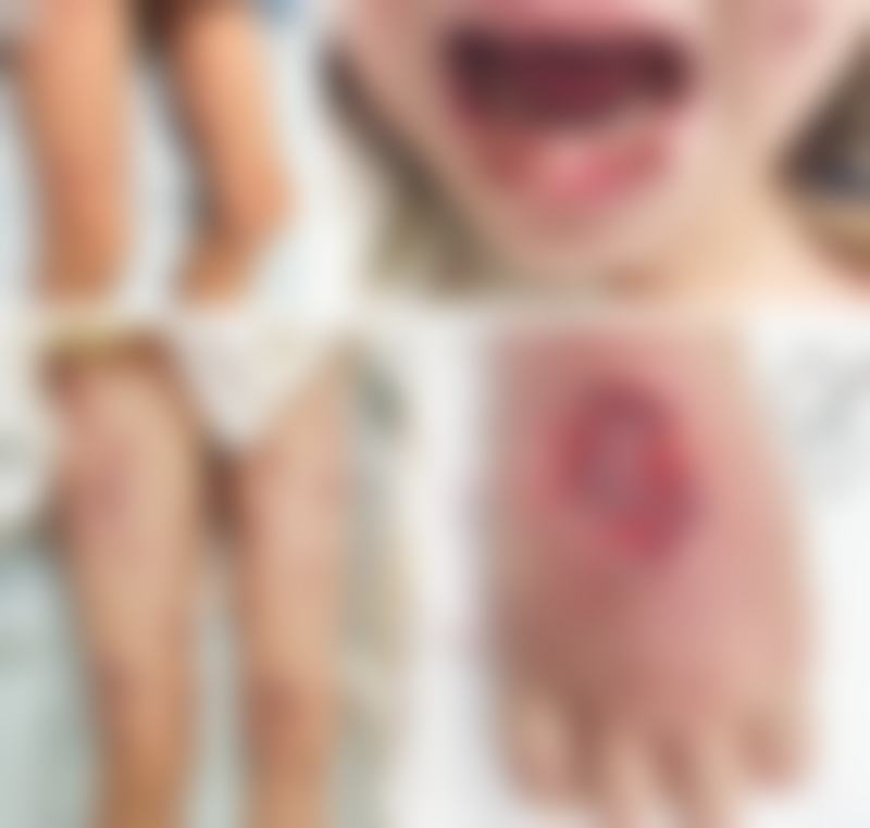
A 7-year-old girl presented to the emergency department with a 2-week history of fever and with several days of painful blisters on her arms, legs, and face. Two weeks earlier, she had completed a course of penicillin for tonsillitis. On examination, there were multiple bullae with erythematous bases on the legs (Panel A) and well-demarcated, painful lesions on the lip and soft palate with a shallow round ulcer on her left cheek (Panel B). Laboratory studies showed a white-cell count of 32,000 per cubic millimeter (reference range, 5000 to 14,500), with 88% neutrophils. Despite treatment with antibiotic agents and acyclovir, the lesions progressed (Panel C).
Tests for infectious causes, including blood cultures, serologic testing for Epstein–Barr virus and Mycoplasma pneumoniae, wound testing for herpes simplex virus and varicella zoster virus, and bacterial culture, were negative. On hospital day 5, a new skin ulcer developed on the right hand at an intravenous cannula site (Panel D). Pathergy, the development of new lesions induced by minor trauma, may be seen in patients with neutrophilic dermatoses such as postinfectious Sweet’s syndrome and bullous pyoderma gangrenosum. A biopsy sample of the ulcer showed a dense neutrophilic infiltrate extending to the base of the lesion, a finding consistent with a neutrophilic dermatosis. The lesions abated with methylprednisolone and oral cyclosporine therapy and wound care. At follow-up 12 months later, most of the lesions had almost completely resolved, with no evidence of ongoing inflammation.

Author
Ji-Hyun Moon, M.B., B.S.
Julie Huynh, B.Biomed.Sci., M.B., B.S.
Children’s Hospital at Westmead, Westmead, NSW, Australia
h.jools@gmail.com