Non-traumatic splenic rupture in a patient with Kasabach–Merritt syndrome
Jitesh Parmar,1 Behnam Shaygi,2 and Mike Nelson1
BMJ Case Rep. 2009; 2009: bcr08.2008.0792.Published online 2009 Mar 5. doi: 10.1136/bcr.08.2008.0792 PMCID: PMC3027749 PMID: 21686627
BACKGROUND
Haemangiomas are vascular lesions resulting from abnormal proliferation of blood vessels. They are the most common paediatric neoplasm.
Kasabach–Merritt syndrome (KMS) is a rare type of vascular lesion with peculiar characteristics. The diagnosis is based upon three basic findings: enlarging haemangioma, thrombocytopenia and consumption coagulopathy. The thrombocytopenia and consumption coagulopathy is known as Kasabach–Merritt phenomenon.1 Almost 200 cases have been reported in the literature since Kasabach and Merritt described the first case in 194020 but there has been only one case report of non-traumatic splenic rupture in a patient with KMS for whom, due to rarity of non-traumatic splenic rupture, splenctomy was not performed, and the patient died of exsanguination.2
Although splenectomy is more hazardous in patients with impaired coagulation, the situation also is more urgent in such patients since exsanguination occurs so rapidly following splenic rupture. Patients with impaired coagulation who develop evidence of splenic haemorrhage should be evaluated quickly and definitively using available imaging. Once splenic haemorrhage has been confirmed, splenectomy should be seriously considered.
The present report describes a patient with KMS who presented with acute abdominal pain, whose non-traumatic splenic rupture was confirmed by CT scan, and he was treated with emergency laparatomy plus splenectomy and blood products administration to control the coagulopathy. The clinical presentation of the case and outcome of selected treatment modalities are discussed in the light of previous studies done in connection with this subject.
CASE PRESENTATION
The patient was a 22-year-old man who was born with misshapen left arm and was later diagnosed with KMS (fig 1). This patient has had multiple treatments including medical, endovascular and even surgical resections of a part of this arteriovenous malformation. The patient had attended hospital and was admitted with acute abdominal pain with diffusely tender abdomen.

Figure 1
Extensive arteriovenous malformation involving whole left upper limb and left hemithorax.
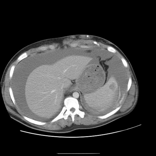
As the patient was haemodynamically stable, and the exact diagnosis was not clear with a background of coagulopathy, an urgent CT scan of the abdomen/chest/pelvis was performed, and this revealed large haemoperitoneum with splenic rupture (fig 2)

INVESTIGATIONS
Investigations on admission were as follows. Full blood count: haemoglobin 6.8 g/dl; mean cell volume 69 fl, platelet count 123×109/l. Clotting: international normalised ratio 1.5, activated partial thromboplastin time ratio 1.59. Cross-match: IgG autoantibody 3+. CT scan of the abdomen/chest/pelvis: large haemoperitoneum with splenic rupture.Go to:
TREATMENT
In view of the CT scan result and coagulopathic blood picture, the patient was resuscitated with blood products and had urgent laparatomy plus splenectomy with successful control of haemorrhage. The patient was then transferred to the intensive treatment unit where he remained unstable. A re-look laparatomy was performed 8 h later, and this revealed no major haemorrhage. Only generalised oozing, especially from splenic bed, was found. This was deemed to be related to on going coagulopathy.
Despite successful control of haemorrhage surgically, the patient continued to remain unstable and required more blood products as follows: 28 units red blood cells, 14 units fresh frozen plasma, 10 units platelets, 10 units cryoprecipitate, and 2× Novo VII. This overwhelmed resources at local district hospital level, and after stabilisation the patient was transferred to a nearby tertiary care unit where he received further 10 units red blood cells, 8 units platelets, 3 units cryoprecipitate, 2 units fresh frozen plasma in the first 24 h.Go to:
OUTCOME AND FOLLOW-UP
The continued coagulopathy was treated with further blood products and systemic corticosteroid therapy. The patient was eventually discharged with full recovery after 4 weeks in hospital.Go to:
DISCUSSION
KMS is a very rare vascular malformation, and more than 80% of cases occur within the first year of life.3 With an overall mortality rate of 12%, there is a mortality rate of 30% for patients with diffuse cavernous haemangiomas when a major haemorrhage occurs.4,5 Various treatment regimens have been published, with inconsistent results. There are two major treatment objectives: the control of the coagulopathy and thrombocytopenia, as well as eradication of the haemangioma. Consequently, different treatment regimens are performed, including systemic corticosteroids,6,7 irradiation,8,9 compression,10 embolisation,11,12 antifibrinolytic agents,13,14 platelet aggregation inhibitors,15 and interferon,16,17 as well as other strategies.18,19
As previously stated there is only one case report in available literature published in 1973: a patient with known KMS presented with spontaneous splenic rupture and unfortunately died because of massive haemorrhage.
LEARNING POINTS
- Although splenectomy is more hazardous in patients with impaired coagulation, the situation also is more urgent in such patients since exsanguination occurs so rapidly following splenic rupture.
- Patients with impaired coagulation who develop evidence of splenic haemorrhage should be evaluated quickly and definitively.
- There are two major treatment objectives: the control of the coagulopathy and thrombocytopenia, as well as eradication of the exsanguination site.
- Once splenic haemorrhage has been confirmed, splenectomy should be seriously considered.
References
1. Abbas AAH, Raddadi AA, Chedid FD.Haemangiomas: a review of the clinical presentations and treatment. Middle East Paediatrics 2003; 8: 52–8 [Google Scholar]2. Stout C, Hampton JW, Anderson JD, et al. Fatal nontraumatic splenic rupture in hemophilia and the Kasabach–Merritt Syndrome. South Med J 1973; 66: 791–5 [PubMed] [Google Scholar]3. Martins AG. Hemangioma and thrombocytopenia. J Pediatr Surg 1970; 5: 641. [PubMed] [Google Scholar]4. El-Dessouky M, Azmy AF, Raine PA, et al. Kasabach–Merritt syndrome. J Pediatr Surg 1988; 23: 109–11 [PubMed] [Google Scholar]5. Camilleri M, Chadwick VS, Hodgson HJF. Vascular anomalies of the gastrointestinal tract. Hepatogastroenterology 1984; 31: 149–53 [PubMed] [Google Scholar]6. Millar JG, Orton CI. Long term follow-up of a case of Kasabach–Merritt syndrome successfully treated with radiotherapy and corticosteroids. Br J Plast Surg 1992; 45: 559–61 [PubMed] [Google Scholar]7. Hagerman LJ, Czapak EE, Donnellan WJ, et al. Giant hemangioma with consumption coagulopathy. J Pediatr 1975; 87: 766–8 [PubMed] [Google Scholar]8. Biswal BM, Anand AK, Aggartwal HN, et al. Vertebral haemangioma presenting as Kasabach–Merritt syndrome. Clin Oncol 1993; 5: 187–8 [PubMed] [Google Scholar]9. Mitsuhashi N, Masaya F, Sakurai H, et al. Outcome of radiation therapy for patients with Kasabach–Merritt syndrome. Int J Radiat Oncol Biol Phys 1997; 39: 467–73 [PubMed] [Google Scholar]10. Aylett SE, Williams AF, Bevan DH, et al. The Kasabach–Merritt syndrome: treatment with intermittent pneumatic compression. Arch Dis Child 1990; 65: 790–1 [PMC free article] [PubMed] [Google Scholar]11. Apfelberg DB, Maser MR, White DN, et al. Combination therapy for massive cavernous hemangioma of the face: YAG laser photocoagulation plus direct steroid injection followed by YAG laser resection with sapphire scalpel tips, aided by superselective embolization. Laser Surg Med 1990; 10: 217–23 [PubMed] [Google Scholar]12. Sato Y, Frey EE, Wicklund B, et al. Embolization therapy in the management of infantile hemangioma with Kasabach–Merritt syndrome. Pediatr Radiol 1987; 17: 503–4 [PubMed] [Google Scholar]13. Warrell RP, Jr, Kempin SJ. Treatment of severe coagulopathy in the Kasabach–Merritt syndrome with aminocaproic acid and cryoprecipitate. N Engl J Med 1985; 313: 309–12 [PubMed] [Google Scholar]14. Neidhart JA, Roach RW. Successful treatment of skeletal hemangioma and Kasabach–Merritt syndrome with aminocaproic acid. Am J Med 1982; 73: 434–8 [PubMed] [Google Scholar]15. Koerper M, Addiego JE, Jr, DE Lorimier AA. Use of aspirin and dipyriadamole in children with platelet trapping syndromes. J Pediatr 1983; 102: 311–4 [PubMed] [Google Scholar]16. Alvarez-Franco M, Paller AS. Congenital Kasabach–Merritt syndrome. Pediatr Dermatol 1994; 11: 80–1 [PubMed] [Google Scholar]17. Hatley RJ, Sabio H, Howell CG, et al. Successful management of an infant with a giant hemangioma of the retroperitoneum and Kasabach–Merritt syndrome with α-interferon. J Pediatr Surg 1993; 28: 1356–9 [PubMed] [Google Scholar]18. Paletta FX, Walker J, King J. Hemangioma-thrombocytopenia syndrome. Plast Reconstruct 1959; 23: 615–20 [PubMed] [Google Scholar]19. Tanaka K, Shimao S, Okada T, et al. Kasabach–Merritt syndrome with disseminated intravascular coagulopathy treated by exchange transfusion and surgical excision. Dermatologica 1986; 173: 90–4 [PubMed] [Google Scholar]20. Hesselmann S, Micke O, Marquardt T, et al. Kasabach–Merritt syndrome: a review of the therapeutic options and a case report of successful treatment with radiotherapy and interferon alpha. Br J Radiol 2002; 75: 180–4 [PubMed] [Google Scholar]
Articles from BMJ Case Reports are provided here courtesy of BMJ Publishing Group
Author Information
1St Mary’s Hospital, General Surgery, Parkhurst Road, Newport, Isle of Wight PO30 5TG, UK2King’s College Hospital, Cardio-Thoracic Surgery, Denmark Hill, London SE5 9RS, UKEmail: moc.oohay@igeyahsb
Copyright and license information
Copyright 2009 BMJ Publishing Group Ltd