Penetrating Heart Injury due to Screwdriver Assault
Case Report
This is a 16-year-old boy who experienced physical aggression in urban fight. He was assaulted by a screwdriver and had stab wound on the anterior face of the chest. He was transported from St. Louis, 192 km away, to our facility by ambulance in a stable hemodynamic status and arrived 8 hours later.
Physical examination showed a screwdriver penetrating the sternum bone in the inferior third over a right angle (Figure 1). Heart bruits were normal. Signs of important bleeding were not seen.

Screwdriver penetrating the chest.
Cardiac ultrasound showed a metallic foreign body in the right ventricle wall with images of thrombus in the right ventricle and a mild pericardial effusion.
The CT scan showed the screwdriver landing into the right ventricle (Figure 2).
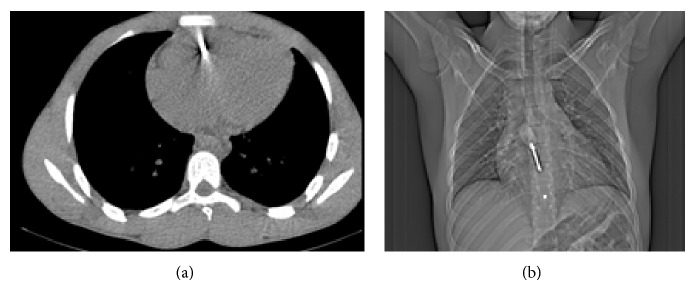
CT scan images of screwdriver inside the heart.
Surgical exploration was done under general anesthesia and orotracheal intubation. Surgical access was a median sternotomy (Figure 3).

Image of screwdriver remaining after sternotomy.
We discovered a mild pericardial blood effusion and a right ventricle wound of 5 mm in diameter with transection of the right coronary vein (Figure 4).

Image of heart stab wound with vein injury after screwdriver removal.
The screwdriver was removed without cardiopulmonary bypass (CPB) and the ventricle wound repaired by direct suture of stitches reinforced with Teflon pledgets. The right coronary vein was ligated.
Postoperative period was free of events. No clinical or biological sign of infection was noted.
Cardiac ultrasound done the day after surgery showed a small thrombus in the lateral wall of the right ventricle. Under heparin therapy, that thrombus disappeared on the 7th day of follow-up.
Author information
Service de Chirurgie Cardiovasculaire et Thoracique, CHUN Fann, Dakar, Senegal
Academic Editor: Monvadi Barbara Srichai
Copyright
Copyright © 2015 P. A. Dieng et al.