Replantation of an Amputated Hand
Case Report
A 46 year old right handed male patient sustained an injury when his dominant hand was caught in a block cutting machine he was working on. He was conscious, oriented, appeared pale and had a BP of 90/60 mm Hg. His right forearm stump showed evidence of crush injury, (Fig 1). The amputated hand was brought in a polythene bag placed inside a plastic box filled with ice. (Figs. 2,3)
Bench surgery was performed on the amputated part by one team while the patient was being prepared for induction. It involved shortening of both bones by about 1.5 cm, debridement of the crushed tissues, and tagging of the arteries, veins and nerves. Ice packs were kept in the vicinity of the part until the vascular continuity was established.
The ulnar and radial arteries were freshened and good bleed was confirmed. The ulnar artery repair was done and good backflow from the distal radial artery was observed. Arterial input was established around 4 hours following the injury. The tourniquet was now elevated and subsequent repairs of the median nerve, ulnar nerve, radial artery, and three veins – two on the dorsum and one on the volar aspect were carried out.
All vascular anastomoses were done with 10-0 nylon under the operating microscope (Zeiss Opmi Vario). The tendons were repaired individually or mass repaired expeditiously to avoid undue delay. The surgery lasted eight hours and the patient recovered from anaesthesia uneventfully.
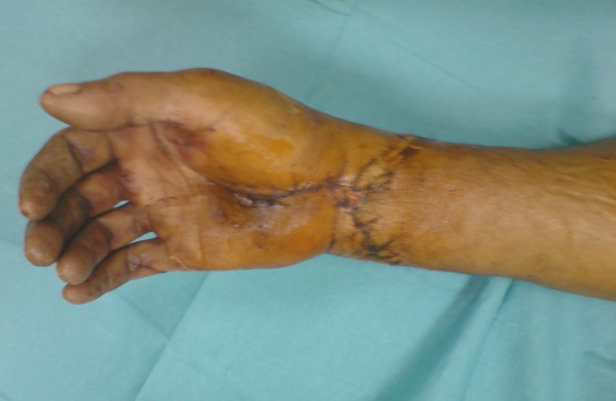
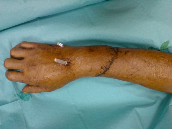
All wounds healed and the patient was discharged after three weeks, (Figs. 4,5).


Credits: Dr. Vipul Nanda, ncbi


