Successful abdominal wall closure following collagen-based artificial dermis induced epithelialization for giant omphalocele: A case report
Masaki Horiike,a,⁎ Tomohiro Kitada,b Kenji Santo,c Takuro Hashimoto,c and Onishi Satoshid
2. Presentation of case
A Japanese female neonate was born at an estimated gestational age of 38 weeks with a birthweight of 3.047 kg. She had an intact GO with a fascial defect measuring 0.06 × 0.06 m (Fig. 1). No other associated abnormalities were found on screening including postnatal echocardiography. G-band analysis showed no chromosomal abnormality and normal karyotype. As the sac containing several protuberant viscera was large, pedunculated, and unsupported, the sac rolled over. The hemodynamics changed and became unstable, leading to low blood pressure.

Fig. 1
Giant omphalocele (GO) findings immediately after birth. A female neonate was born at 38 weeks estimated gestational age with a birthweight of 3047 g. She had a GO with a fascial defect measuring 6 × 6 cm, and no abnormalities were found other than the GO
On the day of birth, a silo was formed using a small size wound retractor (SWR) (Alexis Wound Retractor small size, Applied Medical Resources Corporation, Rancho Santa Margarita, CA, USA) to support the large sac. Since the hepatic veins were firmly adherent to the cranial end of the rectus fascia of the abdominal wall defect, the SWR could not be inserted through the fascial defect. Hence, the SWR was fixed to the abdominal wall (Fig. 2). The silo was crimped daily, but repatriation of the herniated viscera had not progressed. The abdominal wall to which SWR was fixed was incised because numerous injuries had resulted from the excessive tension that had been applied to the fixed parts. Therefore, biomaterial (GORE-TEX® Soft tissue patch, W. L. Gore & Associates, Inc., Newark, NJ, USA) was sutured to each fascia to form a new silo after removing the SWR and sac (Fig. 3). In addition, a sterile dressing of silver (AQUACEL®Ag Extra TM, ConvaTec, England & Wales) was attached to prevent infection in the silo. However, by day 51 the silo had become infected and resulted in bacteremia.

Fig. 2
Findings fixing the small size wound retractor (SWR) to the abdominal wall. Since the hepatic veins firmly adhered to the cranial fascia of the abdominal wall defect and the SWR could not be inserted the fascial defect, the SWR was fixed to the abdominal wall.

Fig. 3
Findings fixing a biomaterial soft tissue patch to the fascia around the abdominal wall defect. A biomaterial soft tissue patch was sutured to the fascia around the abdominal wall defect to form a new silo after removing the small size wound retractor and amniotic membrane.
The infected silo was removed quickly, but a large quantity of pus was noted adhering to the surface of the protruding viscera. A new silo was reformed using an SWR, and the infected surface was washed with saline daily.
By day 65, the infection was under control, and a fibrous capsule had formed on the surface of the viscera and had stabilized them (Fig. 4a). On the other hand, the abdominal wall had become thin and fragile, making it difficult to stretch it and close the abdominal wall. Hence, we decided that it would be impossible to close the wall by reducing the silo contents and considered the idea of epithelializing the abdominal wall defect, leading to closure. We utilized the NPWT method: non-adherent wound dressings (Mepitel®, Molnlycke Healthcare, Gothenburg, Sweden), WHITEFORM® dressings, and GRANUFORM® dressings were applied in this order, and then a clear film was applied to the entire omphalocele. The apex was cut into 1 cm squares and connected to the negative pressure system. The system was set at -25 mmHg (Fig. 4b). Using this method, epithelization proceeded smoothly for a while. On day 71, it was discovered that a part of the epithelializing capsule had ruptured, and the jejunum had perforated (Fig. 4c).

Fig. 4
a–d: Findings from the silo infection control to introducing the negative pressure wound therapy (NPWT) device to the jejunal perforation and taking countermeasures.
Since the new epithelium that had formed was incomplete and fragile, and the perforated jejunum had adhered to the capsule, intestinal fluid did not leak into the abdominal cavity. Thus, the perforated jejunum was managed as a natural intestinal fistula.
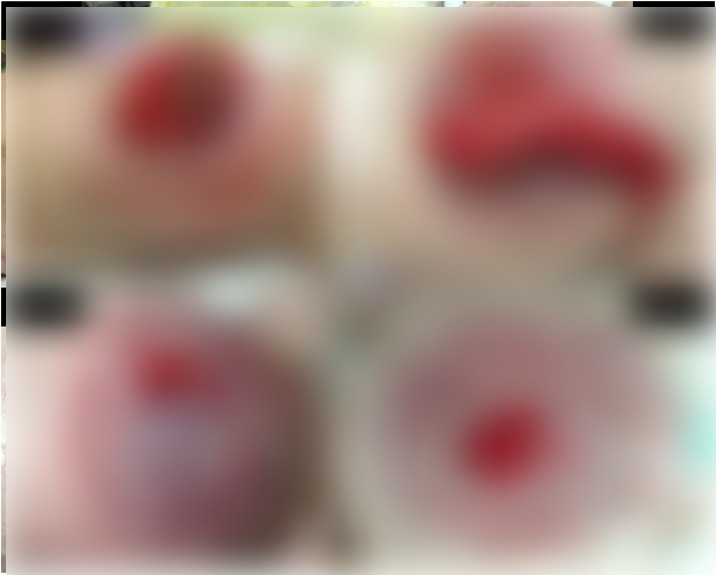
As a result of absorbing the intestinal fluid discharge with gauze, and protecting the epithelial coating with an absorbent foam dressing (Mepilex®, Mölnlycke Health Care AB. Sweden) (Fig. 4d), re-epithelialization of the capsule progressed smoothly (Fig. 5a).

Fig. 5
a–d: Findings from incomplete epithelialization to repairing the perforated jejunum and completing the epithelialization using a collagen-based artificial dermis.
By day 178, the epithelial layer had advanced to the periphery of the natural intestinal fistula (Fig. 5b), and hence, an incision was made along the epithelialization area, and the intestinal fistula was drawn out and repaired. On the other hand, it was impossible to close the incision area with sutures because the abdominal wall was thin and fragile. Therefore, a collagen-based artificial dermis (Terudermis®, Olympus Terumo Biomaterials Corp, Tokyo, Japan) was cut according to the size of the incision area and fixed by suturing (Fig. 5c).
By day 247, epithelialization of the abdominal wall was almost complete (Fig. 5d), and on day 328, the patient was discharged from our hospital. She regularly visits our pediatric surgery outpatient department and was found to be well at the latest follow up, 1 year later, without postoperative complications.
Copyright © 2020 The Author(s)This is an open access article under the CC BY license (http://creativecommons.org/licenses/by/4.0/).