Successful Hand Replantation in a Case of Total Avulsion without Vein Graft
CASE REPORT
A 36-year-old, right-handed male presented to our emergency department after accidentally crushing and severing his hand at the level of the wrist with an industrial roller-type press. The patient applied a bandage to the proximal forearm. The amputated right hand remained in the machine for 2 hours. The patient was taken to the nearest primary health care facility where he was stabilized. The stump was irrigated with 6 L of normal saline, and direct compression was applied to control the bleeding. The stump was wrapped in gauze soaked with normal saline, and a bandage was applied. The severed hand was submerged in saline in a plastic bag and kept in an ice-packed box.
The patient was transferred by ambulance to Riyadh Care Hospital Emergency Department, arriving 6 hours after the initial injury. The warm ischemia time at this point was 2 hours, and the cold ischemia time was 4 hours. The patient arrived alert, oriented, and vitally stable with a blood pressure of 115/80 mm Hg and a heart rate of 96 beats per minute. On examination, the amputated part appeared pale, with multiple-level fractures, the tendons avulse from common flexor and extensor origin, and the vessels and nerves suffered at the level of mid-forearm (Fig.1). The stump was seemed to be physiological degloving injury, contusion wound with skin loss over cubital fossa, contaminated with oil and dust of the machine.

Fig. 1
Intraoperative showing the stump part and amputation part.
The patient was booked for urgent replantation surgery. Preoperative laboratory studies including a complete blood count, cross matching, complete metabolic panel, coagulation profile, and an x-ray of the right hand were ordered (Fig. (Fig.2).2). The patient was started on IV infusion Piperacillin/Tazobactam 4.5 g q8/h for 10 days and oral Clindamycin 450 mg bid for 10 days. The surgery was performed under general anesthesia with an axillary nerve block. The total time of the operation was 7 hours.
The procedure began with the plastic surgeons debriding and exploring the ulnar artery, radial artery, median nerve, and ulnar nerve on both the proximal and distal stumps using vessels loops and clamps. The orthopedic surgeons then proceeded with osteosynthesis, which took 1.5 hours to accomplish. An open reduction and internal fixation of the distal radius and styloid process with size 2.0 K-wires was performed. Next, trans-carpal fixation with a total of six 2.0 K-wires was performed.
Finally, the flexor and extensor tendons were repaired with Prolene 3.0, and the muscles were repaired with Vicryl Rapide 3.0.
The microvascular repairs took 1.5 hours. The vessels were examined under microscopy (10×) to role out intimal injury and access length of segment need to debridement, which was 1.5 mm both ulnar and radial arteries and veins. This involved anastomosis by using Prolene 8.0. Dextran, 3,500 units, was administered intravenously to check the integrity of the anastomosis. After that, the median and ulnar nerves were repaired using Prolene 8.0. A fasciotomy of the forearm flexor compartment was performed to avoid compartment syndrome. Finally, the skin was closed with Vicryl Rapid 3.0 and staples. Bactigras and dressing were applied, and a back slab was cast to immobilize the limb. Secondary closure of skin by split-thickness skin graft was done 10 days later.
Following the surgery, the patient was transferred to the intensive care unit for observation. The patient was kept on heparin, 8000 units/h postoperatively for 7 days. No systemic or major wound complications developed, and the patient was shifted to the surgical ward after 3 days. The patient stayed in the ward for 1 month; he was kept well hydrated in a warm room 80˚F. Pain was well controlled by intravenous 5 mg morphine twice per day and 8 mg IV Lornoxicam twice daily for the first 3 days, then shift it to oral Acetaminophen 500 mg every 8 hours with Lornoxicam 8 mg IV twice daily for 6 days.
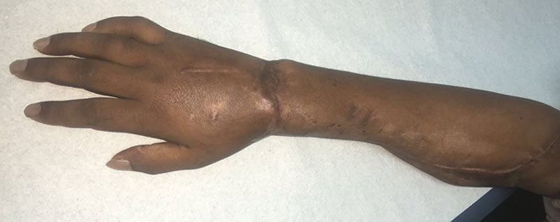
He was managed with daily regular loose dressing. Skin temperature, venous congestion and capillary refill, and pulse oximetry were closely monitored. Physiotherapy was consulted to arrange for rehabilitation sessions. The patient was discharged after 42 days with instructions to stop smoking and follow-up at the clinic with x-rays every 2 weeks to monitor bone healing. The K-wires were removed 2 months later (Fig. 2). One year after the operation, the range of motion active and passive at the wrist was acceptably preserved 45˚ on flexion, 35˚ on extension and 30˚ radioulnar deviation arc. Total active motion was possible for the fingers on flexion and extension, and at the metacarpophalangeal, proximal interphalangeal, distal interphalangeal joints. Grip strength was 5 kg. Sensation was fairly recovered, present along the ulnar nerve distribution, but not in the median nerve or radial nerve distributions. The patient is now able to perform activities of daily living, engage in moderate physical activities.