Thoracic impalement injury: A survivor with large metallic object in-situ
Case report
A 25-year-old male send to emergency department (ED) due to thoracic impalement injury caused by a metallic bar. He crashed into a lorry from behind in high speed when he was driving a motor car. As a result, his car wind screen was broken, a blunt iron bar from the lorry pierced through his upper sternum and came out from posterior aspect of right thorax, below and lateral to tip of right scapula. The patient was stuck at the accident scene for almost an hour with severe pain and mild continuous bleeding. He was separated from damaged vehicle by rescue workers after cutting the metallic bar off the lorry and was brought to hospital with in-situ foreign body. During pre-hospital phase, he received IV fluids resuscitation with 18 g cannula and analgesics.
The patient was conscious when he arrived at emergency room (ER), was having mild tachypnea and agonizing with pain. His pulse was 125 beats per minute, blood pressure (BP) was 100/60 mm Hg, respiratory rate (RR) was 35/min, Glasgow coma scale (GCS) was 15/15 and SpO2 of 92%. Patient was in grade-II hypovolemic shock. There was no past significant medical history. Immediate resuscitation according to advanced trauma life support (ATLS) protocols was conducted. Broad spectrum antibiotics and tetanus immunization was given and right sided tube thoracostomy was done, which caused around 1800 ml fresh blood loss.
When an urgent thoracotomy was planned, regional blood bank was informed that a massive blood transfusion was needed. Without any radiological investigation, the patient was transferred to operating room immediately.
With general anesthesia and double lumen endotracheal tube, patient was put in supine position, slightly towards right edge of operating table to adjust the in-situ rod (Fig. 1, Fig. 2). Complete aseptic measures were applied to the front and back of the patient as well as the metallic rod in order to prevent from any further contamination. Limited right anterolateral thoracotomy was carried out by a team consisted of 2 general surgeons and a cardiothoracic surgeon, also a senior anesthetist who responsible for anesthesia.

Fig. 1 Patient with in-situ object and tube thoracostomy of right chest.

Metallic bar in-situ, also chest tube in place.
We found this rusted metallic rod pierced the chest wall, fractured the adjacent ribs and pierced the right lung parenchyma lead to continuous bleeding. No major bronchial vessel or bronchus was damaged. The bar was removed under vision after separated from lung parenchyma with the help of linear cutting staplers (Fig. 3, Fig. 4). Hemostasis was secured and lung parenchyma damage was repaired.
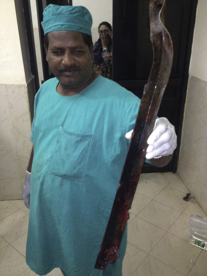

Intraoperative view of foreign body.
In addition, antibiotics was used in operation. Hemostasis of chest wall was obtained. Thoracic cavity was lavaged with 2 L of warm normal saline. Chest wall closed with non-absorbable prolene-1 sutures and muscles repaired with absorbable Vicryl-1 sutures. Skin closed with skin staplers. With a chest drain left in place, the patient was transferred to ICU, where he had kept for 3 days.
Post-operative chest X-ray showed fully expanded lung with no collection. Chest tube was removed at 4th day after operation. Patient was discharged at 5th day after operation. His follow-up visits were unremarkable and no significant sequelae of the injury were seen.
Written informed consent was obtained from the patient for publication of this case report and accompanying images. A copy of the written consent is available for review by the Editor-in-Chief of this journal on request.
References
- WHO . 2013. Global Status Report on Road Safety 2013: Supporting a Decade of Action. Geneva, Switzerland. [Google Scholar]
- Yates D.W., Woodford M., Hollis S. Preliminary analysis of the care of injured patients in 33 British hospitals: first report of the United Kingdom major trauma outcome study. BMJ. 1992;305:737–740. [PMC free article] [PubMed] [Google Scholar]
- Hyde M.R., Schmidt C.A., Jacobson J.G. Impalement injuries to the thorax as a result of motor vehicle accidents. Ann Thorac Surg. 1987;43:189–190. [PubMed] [Google Scholar]
- Sawhney C., D’Souza N., Mishra B. Management of a massive thoraco-abdominal impalement: a case report. Scand J Trauma Resusc Emerg Med. 2012;18 57-57. [Google Scholar]
- Bemelman M., Hammacher E. Rectal impalement by pirate ship: a case report. Inj Extra. 2005;36:508–510. [Google Scholar]
- Oya S., Miyata K., Yuasa N. Impalement injury to the left buttock with massive bleeding: a case report. Nagoya J Med Sci. 2013;75:147–152. [PMC free article] [PubMed] [Google Scholar]
- Angelopoulos S., Ioannis M., Dimitrios K. A rare case of a transabdominal impalement after a fall from a ladder. Int J Surg Case Rep. 2016;22:40–43. [PMC free article] [PubMed] [Google Scholar]
- Robicsek F., Daugherty H.K., Stanfield A.V. Massive chest trauma due to impalement. J Thorac Cardiovasc Surg. 1984;87:684–686. [PubMed] [Google Scholar]
- Chui W.H., Cheung D.L., Chiu S.W. A non-fatal impalement injury of the thorax. J R Coll Surg Edinb. 1998;43:419–421. [PubMed] [Google Scholar]
- Cartwright A.J., Taams K.O., Unsworth-White M.J. Suicidal nonfatal impalement injury of the thorax. Ann Thorac Surg. 2001;72:1364–1366. [PubMed] [Google Scholar]
- Shimokawa S., Shiota K., Ogata S. Impalement injury of the thorax:report of a case. Surg Today. 1994;24:926–928. [PubMed] [Google Scholar]
- Wood A.E. Transfixion injury of the chest. J Trauma. 1982;22:432–433. [PubMed] [Google Scholar]
- Thomson B.N., Knight S.R. Bilateral thoracoabdominal impalement: avoiding pitfalls in the management of impalement injuries. J Trauma. 2000;49:1135–1137. [PubMed] [Google Scholar]
- Darbari A., Tandon S., Singh A.K. Thoracic impalement injuries. Ind J Thorac Cardiovasc Surg. 2005;21:229–231. [Google Scholar]
Articles from Chinese Journal of Traumatology are provided here courtesy of Elsevier
Original Source
Copyright and License information
Copyright © 2018 Daping Hospital and the Research Institute of Surgery of the Third Military Medical University. Production and hosting by Elsevier B.V.This is an open access article under the CC BY-NC-ND license (http://creativecommons.org/licenses/by-nc-nd/4.0/).