Dermoscopy of Pigmented Bowen’s Disease Mimicking Early Superficial Spreading Melanoma
Yuka Hayashi
aDepartment of Dermatology, Hiratsuka City Hospital, Hiratsuka, Japan
Masaru Tanaka
bTokyo Women’s Medical University Medical Center East, Tokyo, Japan
Reiko Suzaki
bTokyo Women’s Medical University Medical Center East, Tokyo, Japan
Nuiko Mori
aDepartment of Dermatology, Hiratsuka City Hospital, Hiratsuka, Japan
Izumi Konohana
aDepartment of Dermatology, Hiratsuka City Hospital, Hiratsuka, Japan
Abstract
A 89-year-old Japanese woman presented at our clinic because of a several months’ history of an asymptomatic gradually enlarging pigmented skin lesion on the dorsum of the left foot. Physical examination revealed a single hyperpigmented oval macule of 5 mm with a rough surface. The color of the lesion was dark brown to light brown. Dermoscopic examination demonstrated atypical pigment network with small dotted vessels. Irregular streaks were also partially noted at the periphery. We suspected superficial spreading melanoma and performed an excision. The histologic features were consistent with a diagnosis of pigmented Bowen’s disease. We could not completely account for dermoscopic aspects from the pathological findings of hematoxylin and eosin-stained specimens; therefore, specimens were stained with Fontana-Masson stain. It clearly demonstrated the distribution of melanin in the epidermis. We concluded that atypical network was due to an uneven melanin deposition in the variably thickened epidermal rete ridges.
Case Report
A 89-year-old Japanese woman presented at our clinic because of a several months’ history of an asymptomatic gradually enlarging pigmented skin lesion on the dorsum of the left foot, which had been 1 mm at the beginning. Physical examination revealed a single hyperpigmented oval macule of 5 mm with a rough surface. The color of the lesion was dark brown to light brown. The lesion was slightly elevated (fig. 1). Dermoscopic examination demonstrated an atypical pigment network with small dotted vessels (fig. 2). Irregular flossy streaks were also partially noted at the periphery. We suspected very early superficial spreading melanoma and performed an excision with a 3 mm margin.
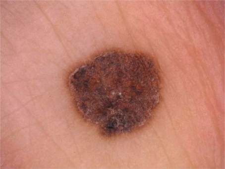
A pigmented macule of 5 mm on the dorsum of the left foot.

Dermoscopy demonstrates atypical pigment network and dotted vessels. Irregular flossy streaks are also partially noted.
Histopathological examination of the hematoxylin and eosin-stained specimens disclosed the following (fig. 3): the epidermis showed acanthosis with elongation and thickening of the rete ridges. In the upper epidermis, prominent vacuolated cells were observed. The border between the epidermis and the dermis appeared sharp. Atypical multinucleate keratinocytes, dyskeratotic cells, and mitoses were seen in all the layers of the epidermis. The stratum corneum showed hyperkeratosis and parakeratosis without the granular layer. Melanin pigment was irregularly observed throughout the epidermis including the stratum corneum. The basal melanosis was also irregularly distributed. Melanophages were occasionally noted in the papillary dermis. The histologic features were consistent with a diagnosis of pigmented Bowen’s disease.

Hematoxylin and eosin-staining shows hyperkeratosis with parakeratosis, vacuolar changes in the upper portion of the epidermis and proliferation of atypical keratinocytes throughout the epidermis.
Although melanin pigment seemed to be present in the whole epidermal layer, it would not fully explain the feature of dermoscopy showing atypical pigment network. Therefore, we also performed Fontana-Masson staining, which revealed that melanin pigment covered a wider area, including the basal layer, and visualized its irregular distribution more clearly than the hematoxylin and eosin-stained specimens (fig. 4).

Fontana-Masson stain discloses uneven distribution of melanin in the epidermis.
Discussion
Pigmented Bowen’s disease is rare and presents clinically as a pigmented macule with a scaly or verrucous surface. Therefore, seborrhoeic keratosis, pigmented actinic keratosis, solar lentigo, basal cell carcinoma and melanoma in situ should occasionally be included in the differential diagnosis.
Dermoscopic examination of Bowen’s disease generally demonstrates glomerular vessels and scaly surface. There are only a few reports about pigmented Bowen’s disease. Zalaudek et al. reported that pigmented Bowen’s disease showed dermoscopic features such as small brown globules, grey to brown homogeneous pigmentation, and they also depicted that pigment network and streaks were seen in heavily pigmented Bowen’s disease [1]. Stante et al. also published a report on pigmented Bowen’s disease which showed atypical pigment network on dermoscopy like our case, and they also commented that cutaneous melanoma could not be denied [2].
Pigment network is considered one of the most specific parameters of melanocytic proliferation and is defined as a subtle network of brownish lines over a diffuse background. It correlates with the presence of melanin within the basal layer of the epidermis. A typical network is generally marked by a uniform deposition of melanin at the basal layer and a diminution of it at the upper epidermis. The lack of balance of melanin deposition at the basal layer forms atypical pigment network [2].
In our case, we could not completely account for dermoscopic aspects, such as atypical pigment network, from the pathological findings of hematoxylin and eosin-stained specimens; therefore, specimens were stained with Fontana-Masson stain. It clearly demonstrated the distribution of melanin in the epidermis, which was not uniform. We concluded that the atypical network in this case was due to an uneven melanin deposition in the variably thickened epidermal rete ridges.
An observation of irregular flossy streaks has never been reported before, but it may be one of the characteristic features of pigmented Bowen’s disease. Therefore, we would like to prompt similar reports. Dermoscopy is considered as a helpful tool for increasing diagnostic accuracy of histopathologic diagnosis of pigmented skin lesion. However, unfortunately in our case, dermoscopic evaluation of the lesion was not helpful in order to reach a correct preoperative classification. When evaluating a pigmented skin lesion like our patient’s by means of dermoscopy, pigmented Bowen’s disease, an unusual epithelial tumor, should also be included in the differential diagnosis. Then, the pathological examination would finally lead to the correct diagnosis.
References
Content retrieved from: https://www.ncbi.nlm.nih.gov/pmc/articles/PMC2895203/.