Giant recurrent lipoma of trunk weighing eight kilograms
A lot of the diseases found on this site, are preventable. Please make sure you’re regular on your check ups and also make sure your healthcare program has you completely covered. If you’re unsure if your selected healthcare plan covers any disease or want more information, you can call for free at +1-877-759-8006, where you will be connected to a healthcare center with an agent on the line. Clicking the number will redirect you to your default call making application.
Case report
An 82-year old male patient presented with a massive subcutaneous swelling extending over the left trunk. He gave history of 4 previous attempts at removal with recurrences within 1–2 years. He was resigned to his fate till the massive proportions interfered with his limb function and inability to lie flat.
Examination revealed a healthy male with scoliosis and obvious dropping of left shoulder. There was a massive multilobulated swelling extending from the left shoulder and back to the posterior arm, axilla and across to the anterior chest. It occupied the entire left posterior trunk from shoulder till iliac crest and was indiscrete around the axilla. Overlying skin had healed scars of previous surgery with dilated veins (Fig. 1). Shoulder movement was restricted due to mass effect and weight. There was no neurovascular deficit or edema noted in the left upper limb. Two additional medium size lipomas were noted at the nape of neck and rt shoulder.
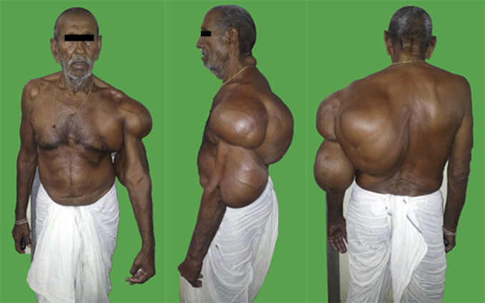
Giant recurrent lipoma extending across left arm, shoulder, chest, axilla and posterior trunk. Well healed faint scars of previous 4 surgeries are seen.
FNAC from multiple sites revealed a lipoma. MRI demonstrated extensive submuscular extension under the pectorals, deltoid, trapezius and latissmus dorsi muscle with no involvement of axillary neurovascular structures (Fig. 2).
The patient had no comorbidities and was operated under general anesthesia in a semiprone position. A 45 cm incision was made commencing from the posterior rt elbow, arm, shoulder and through the lateral back to the iliac crest. The lipoma was dissected starting from the elbow upward. Apart from the subcutaneous portion there was extensive tumor under the deltoid with insinuation under the coraco acromium. It had also spread under the pectorals, trapezius and latissmus across the axilla, from where it was dissected safeguarding the neurovascular structures. Submuscular portions were removed after splitting the overlying muscles in a function preserving approach (Fig. 3). The deltoid was thinned out to less than a cm thickness.

Fig. 3
Intraoperative muscle split approach to deliver the sublatissmus component.
There was moderate blood loss of 700 ml. The entire lipoma was contiguous with a discernable capsule. The removed specimen weighed 8000 g and measured 106 × 18 cm (Fig. 4). Redundant skin was excised and closure done over suction drains. The patient had an uneventful recovery with drain removal on 4th post-operative day. There was no collection, skin necrosis or wound healing problem. After thorough grossing, histopathological examination of multiple sections revealed no evidence of necrosis or malignancy. Early follow up at 3 months did not reveal any evidence of recurrence on clinical exam and Ultrasonography. He has been advised regular follow up at 6 monthly intervals till a year.

Fig. 4
The excised specimen. It weighed 8 kg and measured 106 × 18 cm.
Authors
aAssociate Professor, Dept of Surgery, Armed Forces Medical College, Pune 411040, India
bAssociate Professor, Dept of Radiology, Armed Forces Medical College, Pune 411040, India
cClassified Specialist (Pathology), Command Hospital (Southern Command), Pune 411040, India
Sandeep Mehrotra:
References
References
- Vandeweyer E., Scagnol I. Axillary giant lipoma: a case report. Acta Chir Belg. 2005;105:656–657. [PubMed] [Google Scholar]
- Sanchez M.R., Golomb F.M., Moy J.A., Potozkin J.R. Giant lipoma: case report and review of the literature. J Am Acad Dermatol. 1993;28:266–268. [PubMed] [Google Scholar]
- Nakamura Y., Fujisawa Y., Obara S. Giant lipoma with fat necrosis of the back mimicking atypical lipomatous tumor in MRI findings. J Clin Exp Dermatol Res. 2013;S6:013. [Google Scholar]
- Leuzzia G., Cesarioa A., Parisia A.M., Granonea P. Chest wall giant lipoma with a thirty-year history. Interact Cardiovasc Thorac Surg. 2012;15:323–324. [PMC free article] [PubMed] [Google Scholar]
- Basmani M., Hasturk A.E. Giant occipitocervical lipomas: evaluation with two cases. J Cutan Aesthet Surg. 2012;5:207–209. [PMC free article] [PubMed] [Google Scholar]
- Sultan M.M. Recurrent Fibrolipoma of the left thigh – a case report. Gen Surg. 2012;3 Webmed Central. WMC003378. [Google Scholar]
- Silistreli O.K., Durmuş E.U., Ulusal B.G. What should be the treatment modality in giant cutaneous lipomas? Review of the literature and report of 4 cases. Br J Plast Surg. 2005;58:394–398. [PubMed] [Google Scholar]
- Verma S., Varma M., Kala S., Singh R.K. Giant lipoma of posterior neck with bleeding decubitus ulcer: a rare entity. J Cutan Aesthet Surg. 2010;3:119–121. [PMC free article] [PubMed] [Google Scholar]
Articles from Medical Journal, Armed Forces India are provided here courtesy of Elsevier