Patient with penetrated Wooden Stick
Case Report
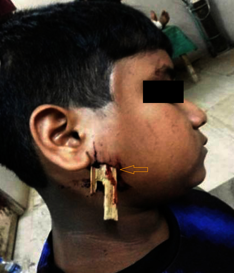
A 15-year-old boy reported to emergency department with history of fall from a tree onto a wooden fence and sustained a penetrating injury from a wooden piece [Fig-1]. There was no evidence of facial nerve injury. CT scan revealed that the foreign body was superficial to the investing layer of deep cervical fascia and all the major neurovascular structures were intact. The foreign body was visualized as linear hypo dense structure situated in the plane superficial to parotid fascia extending cranially superficial to temporal fascia [Fig-2]. After identification of the retained foreign object [Fig-3], surgical exploration and removal of the foreign body in Toto, precisely to prevent neurovascular damage under general anaesthesia was planned.
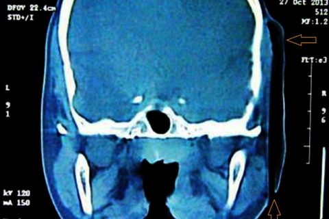
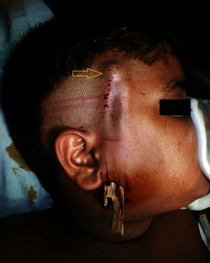
Cranial extension of the foreign body.
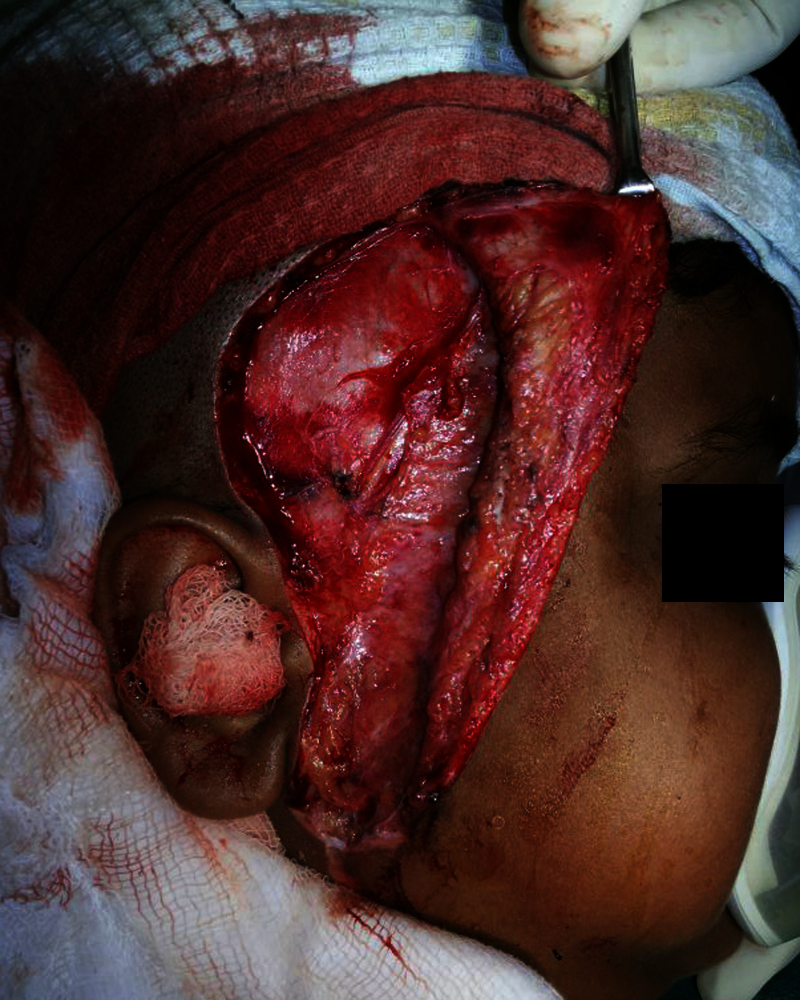
Flap was reflected in a uniform plane to completely expose the wooden piece [Fig-4]. Foreign body was passively extracted and debridement was done to remove any debris [Fig-5]. The local area was irrigated with antiseptic solution, and tissue approximation was completed in layers. The incision was closed with simple interrupted sutures [Fig-6]. The retrieved foreign body was a bamboo stick which measured 11×2 cm [Fig-7].
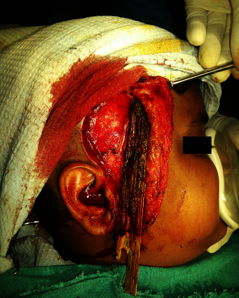
Flap raised for exploration of the site.
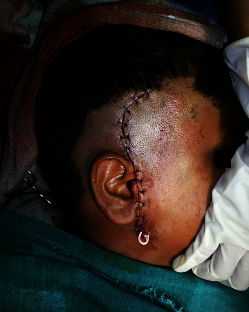
Wound closure and placement of glove drain.
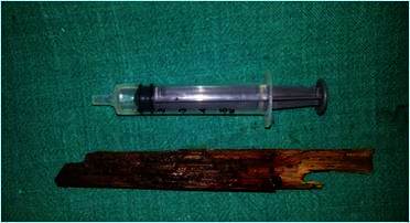
Retrieved foreign body measuring 11x 2 cm.
Patient with stood the procedure and there was no bleeding after the surgery. On the second postoperative day evaluation for facial nerve injury and parotid duct injury was done. Patient was discharged after one week of surgery.
Patient was under follow up for 6 months at regular intervals. In conclusion, this was a rare presentation of penetrating injury without injuring major neck vessels and facial nerve.
Credits: Ncbi, © 2016 Journal of Clinical and Diagnostic Research