Trauma from car accident
Case Report
A twenty-three-year old male patient was brought to our emergency service by an ambulance. Trauma mechanism reported by the paramedic team indicated that the patient was seated in the driver seat without his seatbelt on, and he had crushed his face to the steering wheel during the accident

Sensitive image, click to unblur and zoom
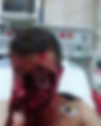
Figure 1
Airway compromise should occur due to tongue falling back, hemorrhage to oropharyngeal region, foreign bodies and mid facial fractures themselves. In this case, it’s hard to define proper tracheal structure to perform endotracheal intubation because of severe mid face fractures.
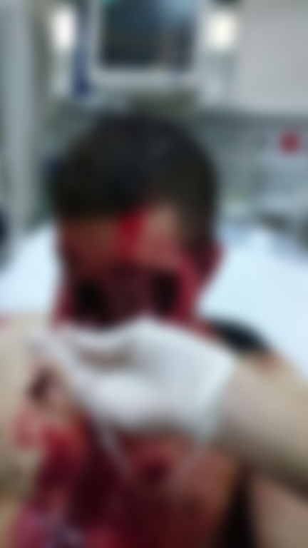
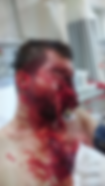
After the initial examination, it was noted that the patient’s bilateral orbitas; maxillary, zygomatic and nasal bone structures were not observed. The patient was conscious, breathing spontaneously in tripod position and he had tachypnea. He had midfacial bleeding due to maxillofacial trauma. He was cooperated and oriented but vocal and ocular responses were suboptimal due to damages. Vital signs were: blood pressure 160/90 mmHg, heart rate 110, respiratory rate 22 and fingertip saturation 99%.(Figure1, figure 2, Figure 3)
On his primary survey, his hemorrhage control was maintained with aspiration. Oxygen saturation was normal while sitting but he was not able to keep his airway open in supine position. After stabilization of the patient, urgent plastic surgery was planned. Because of his severe facial fractures, surgical airway (tracheostomy) placement was performed accompanied by a otolaryngologist. On his secondary survey, other system examinations were normal.
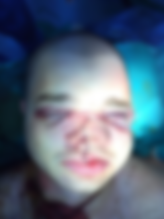
In his cranial computed tomography (CT) imaging there was neither damage in brain parenchyma; nor hemorrhage or hematoma on epidural, subdural or subarachoidal spaces. Lateral, superior, medial and inferior walls of bilateral orbitas and bilateral multiple zygomatic arch fractures were detected. Right bulbus oculi was not in its cavity, left bulbus oculi was displaced antero-superiorly. The relationship between the left temporomandibular joint was absent. After performing cranial imaging, the patient was referred to Plastic and Reconstructive Surgery for operation (figure 4).

Sensitive image, click to unblur and zoom
Figure 2
In most cases, bleeding can be are easily controlled. Rarely, Facial trauma arises from the maxillary artery, creating difficulty in hemorrhage control. In this case suction provided adequate hemorrahge control.

