The Big Bang: Facial Trauma Caused by Recreational Fireworks
Authors: Josher Molendijk, BSc, MSc,1 Bob Vervloet, BSc, MSc,1 Eppo B. Wolvius, DDS, MD, PhD,1 and Maarten J. Koudstaal, MD, DDS, PhD1
Case
A 29-year-old man was hit directly in the face by a rocket in the night of January 1, 2011. The patient was seen at the emergency department of our institution according to the Advanced Trauma Life Support protocol. His airway was threatened due to serious bleeding from extensive soft-tissue injury and the patient was immediately intubated with an endotracheal tube, under the use of etomidate as sedative and succinylcholine as paralytic agent. The patient was hemodynamically stable. Severe soft-tissue injuries to the right side of the face were observed (Fig. 1). Adequate examination of vision and sensibility of the face could not be performed. Computed tomography (CT) angiography images of the face, cranium, and the neck were obtained and revealed multiple fractures as follows: communitive fracture of the mandible; a Le Fort 2 fracture; a midline fracture of the maxilla; extensive fractures of the walls of the right maxillary sinus and both right and left orbital floor; a right zygomatic arch fracture; and fracture of the right temporal bone at the temporomandibular joint. Also noted were small hyperdense, spherical bodies, which correspond with remnants of explosive powder. Angiography revealed an arterial bleeding on the right side of the face. No intracranial or cervical injuries were found.

Fig. 1
Severe facial soft-tissue injury after firework blast to the face in patient 1.
Oral and maxillofacial surgery, ophthalmology, and plastic surgery services were consulted and the patient was brought to the operating room immediately after presentation. Extensive debridement by the plastic surgeons was performed and large defects of facial musculature in the buccal region and lesions of the facial nerve and parotid duct were encountered. The facial artery was damaged and clipped intraoperatively. Much explosive powder residue was removed. After replacement of the oral endotracheal tube with a nasopharyngeal tube, the oral and maxillofacial surgeons approached the fractures of the mandible and maxilla through oral incisions. The fractures were reduced and fixated with titanium plates. Finally, the parotic duct was sutured back into the buccal mucosa and the soft tissue was closed.
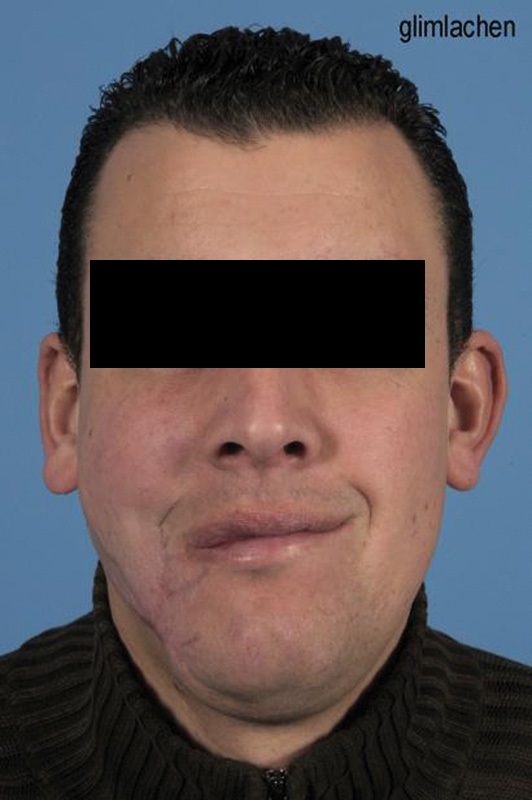
Clinical photograph of patient 1 after the reconstructive surgeries. Drooping of the right corner of the mouth is seen due to facial nerve injury.
The patient was brought to the intensive care for observation. The patient was discharged in good clinical condition on hospital day 9. He was readmitted on day 14 posttrauma, after developing a wound dehiscence in the right buccal region, with formation of an orofacial fistula. The plastic surgeon closed the defect with a supraclavicular artery island flap 33 days posttrauma. At 16 and 20 months posttrauma, multiple scar corrections were performed, to correct mutilating scars in the face and a trismus. Healing of the fractures was uncomplicated and vision was only slightly affected. Drooping of the right corner of the mouth persisted due to facial nerve injury, but did not bother the patient (Fig. 2).
References
1. Keunen J EE, Storimans C WJM. With an eye on… the champagne cork [in Dutch] Ned Tijdschr Geneeskd. 1994;138(52):2594–2596. [PubMed] [Google Scholar]2. Puri V, Mahendru S, Rana R, Deshpande M. Firework injuries: a ten-year study. J Plast Reconstr Aesthet Surg. 2009;62(9):1103–1111. [PubMed] [Google Scholar]3. Veiligheid N L Vuurwerkongevallen 2013–2014. Amsterdam Available at: http://www.veiligheid.nl/csi/veiligheidnl.nsf/0/D5008D18820C51E3C1257C7E004D7191/$file/Vuurwerkongevallen%202013-2014.pdf. Accessed January 10, 20154. Veiligheid N L Beperking afsteektijden zorgt voor minder vuurwerkslachtoffers. Amsterdam http://www.veiligheid.nl/nieuws/beperking-afsteektijden-zorgt-voor-minder-vuurwerkslachtoffers. Accessed January 10, 20155. Edskes S N, Smeulders M JC, van der Zee C W, Zophel O T, Van de Kar A L. Jaarwisseling 2012–2013: Vuurwerkletsels behandeld door plastisch chirurgen. Ned Tijdschr Plastische Chirurgie. 2014;5:63–65. [Google Scholar]6. de Faber J THN. Fireworks injuries treated by Dutch ophthalmologists New Year 2008/’09 [in Dutch] Ned Tijdschr Geneeskd. 2009;153:A507. [PubMed] [Google Scholar]7. Wisse R P, Bijlsma W R, Stilma J S. Ocular firework trauma: a systematic review on incidence, severity, outcome and prevention. Br J Ophthalmol. 2010;94(12):1586–1591. [PubMed] [Google Scholar]8. Romano F, Catalfamo L, Siniscalchi E N. et al.Complex craniofacial trauma resulting from fireworks blast. J Craniofac Surg. 2008;19(2):322–327. [PubMed] [Google Scholar]9. Di Benedetto G, Grassetti L, Forlini W, Bertani A. An explosion in the mouth caused by a firework. J Plast Reconstr Aesthet Surg. 2009;62(6):e145–e146. [PubMed] [Google Scholar]10. Tadisina K K, Abcarian A, Omi E. Facial firework injury: a case series. West J Emerg Med. 2014;15(4):387–393. [PMC free article] [PubMed] [Google Scholar]11. Nam S M. An explosion in the oral cavity by a firecracker. J Craniofac Surg. 2013;24(5):e510–e512. [PubMed] [Google Scholar]12. Weichel E D, Colyer M H, Ludlow S E, Bower K S, Eiseman A S. Combat ocular trauma visual outcomes during operations Iraqi and enduring freedom. Ophthalmology. 2008;115(12):2235–2245. [PubMed] [Google Scholar]13. NOS Vuurwerk heeft kracht handgranaat Available at: http://nos.nl/artikel/590986-vuurwerk-heeft-kracht-handgranaat.html. Accessed January 15, 2015
Articles from Craniomaxillofacial Trauma & Reconstruction are provided here courtesy of SAGE Publications